
Moesin Rabbit mAb [xMN9]Cat NO.: A73963
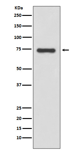
Western blot(SDS PAGE) analysis of extracts from HeLa cell lysate.Using Moesin Rabbit mAb [xMN9]at dilution of 1:1000 incubated at 4℃ over night.
Product information
Protein names :MSN; Moesin;
UniProtID :P26038
MASS(da) :67,820
MW(kDa) :68kDa
Form :Liquid
Purification :Affinity-chromatography
Host :Rabbit
Isotype : IgG
sensitivity :Endogenous
Reactivity :Human,Mouse,Rat
- ApplicationDilution
- 免疫印迹(WB)1:1000-2000
- 免疫组化(IHC)1:100
- 免疫荧光(ICC/IF)1:100
- The optimal dilutions should be determined by the end user
Specificity :Antibody is produced by immunizing animals with A synthesized peptide derived from human Moesin
Storage :Antibody store in 10 mM PBS, 0.5mg/ml BSA, 50% glycerol. Shipped at 4°C. Store at-20°C or -80°C. Products are valid for one natural year of receipt.Avoid repeated freeze / thaw cycles.
WB Positive detected :HeLa cell lysate.
Function : Ezrin-radixin-moesin (ERM) family protein that connects the actin cytoskeleton to the plasma membrane and thereby regulates the structure and function of specific domains of the cell cortex. Tethers actin filaments by oscillating between a resting and an activated state providing transient interactions between moesin and the actin cytoskeleton (PubMed:10212266). Once phosphorylated on its C-terminal threonine, moesin is activated leading to interaction with F-actin and cytoskeletal rearrangement (PubMed:10212266). These rearrangements regulate many cellular processes, including cell shape determination, membrane transport, and signal transduction (PubMed:12387735, PubMed:15039356). The role of moesin is particularly important in immunity acting on both T and B-cells homeostasis and self-tolerance, regulating lymphocyte egress from lymphoid organs (PubMed:9298994, PubMed:9616160). Modulates phagolysosomal biogenesis in macrophages (By similarity). Participates also in immunologic synapse formation (PubMed:27405666)..
Tissue specificity :In all tissues and cultured cells studied.
Subcellular locationi :Cell membrane,Peripheral membrane protein,Cytoplasmic side. Cytoplasm, cytoskeleton. Apical cell membrane,Peripheral membrane protein,Cytoplasmic side. Cell projection, microvillus membrane,Peripheral membrane protein,Cytoplasmic side. Cell projection, microvillus.
IMPORTANT: For western blots, incubate membrane with diluted primary antibody in 1% w/v BSA, 1X TBST at 4°C overnight.
-
About
-
Product
-
Law
-
Literature
-

WeChat scan code follow us