
CD68 Rabbit mAb [hM6g]Cat NO.: A60801
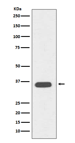
Western blot(SDS PAGE) analysis of extracts from Jurkat cell lysate.Using CD68 Rabbit mAb [hM6g]at dilution of 1:1000 incubated at 4℃ over night.
Product information
Protein names :CD68 antigen; CD68 molecule; Gp110; LAMP4; Macrophage antigen CD68 (microsialin); Macrosialin; SCARD1; Scavenger receptor class D member 1;
UniProtID :P34810
MASS(da) :37,408
MW(kDa) :37kDa
Form :Liquid
Purification :Affinity-chromatography
Host :Rabbit
Isotype : IgG
sensitivity :Endogenous
Reactivity :Human,Mouse,Rat
- ApplicationDilution
- 免疫印迹(WB)1:1000-2000
- The optimal dilutions should be determined by the end user
Specificity :Antibody is produced by immunizing animals with A synthesized peptide derived from human CD68
Storage :Antibody store in 10 mM PBS, 0.5mg/ml BSA, 50% glycerol. Shipped at 4°C. Store at-20°C or -80°C. Products are valid for one natural year of receipt.Avoid repeated freeze / thaw cycles.
WB Positive detected :Jurkat cell lysate.
Function : Could play a role in phagocytic activities of tissue macrophages, both in intracellular lysosomal metabolism and extracellular cell-cell and cell-pathogen interactions. Binds to tissue- and organ-specific lectins or selectins, allowing homing of macrophage subsets to particular sites. Rapid recirculation of CD68 from endosomes and lysosomes to the plasma membrane may allow macrophages to crawl over selectin-bearing substrates or other cells.
Tissue specificity :Highly expressed by blood monocytes and tissue macrophages. Also expressed in lymphocytes, fibroblasts and endothelial cells. Expressed in many tumor cell lines which could allow them to attach to selectins on vascular endothelium, facilitating their dissemination to secondary sites..
Subcellular locationi :[Isoform Short]: Cell membrane,Single-pass type I membrane protein.,[Isoform Long]: Endosome membrane,Single-pass type I membrane protein. Lysosome membrane,Single-pass type I membrane protein.
IMPORTANT: For western blots, incubate membrane with diluted primary antibody in 1% w/v BSA, 1X TBST at 4°C overnight.
-
About
-
Product
-
Law
-
Literature
-

WeChat scan code follow us