
CDK8 Rabbit mAb [Ir21]Cat NO.: A12474
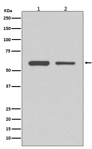
Western blot(SDS PAGE) analysis of extracts from (1) HeLa cell lysate; (2) 3T3 cell lysate.Using CDK8 Rabbit mAb [Ir21]at dilution of 1:1000 incubated at 4℃ over night.
Product information
Protein names :CDK8 protein kinase; Cell division protein kinase 8; Cyclin Dependent kinase 8; K35; Mediator complex subunit cdk8; Mediator of RNA polymerase II transcription subunit cdk8; Protein kinase K35;
UniProtID :P49336
MASS(da) :53,284
MW(kDa) :53kDa
Form :Liquid
Purification :Affinity-chromatography
Host :Rabbit
Isotype : IgG
sensitivity :Endogenous
Reactivity :Human,Mouse,Rat
- ApplicationDilution
- 免疫印迹(WB)1:1000-2000
- The optimal dilutions should be determined by the end user
Specificity :Antibody is produced by immunizing animals with A synthesized peptide derived from human CDK8
Storage :Antibody store in 10 mM PBS, 0.5mg/ml BSA, 50% glycerol. Shipped at 4°C. Store at-20°C or -80°C. Products are valid for one natural year of receipt.Avoid repeated freeze / thaw cycles.
WB Positive detected :(1) HeLa cell lysate; (2) 3T3 cell lysate.
Function : Component of the Mediator complex, a coactivator involved in regulated gene transcription of nearly all RNA polymerase II-dependent genes. Mediator functions as a bridge to convey information from gene-specific regulatory proteins to the basal RNA polymerase II transcription machinery. Mediator is recruited to promoters by direct interactions with regulatory proteins and serves as a scaffold for the assembly of a functional pre-initiation complex with RNA polymerase II and the general transcription factors. Phosphorylates the CTD (C-terminal domain) of the large subunit of RNA polymerase II (RNAp II), which may inhibit the formation of a transcription initiation complex. Phosphorylates CCNH leading to down-regulation of the TFIIH complex and transcriptional repression. Recruited through interaction with MAML1 to hyperphosphorylate the intracellular domain of NOTCH, leading to its degradation..
Subcellular locationi :Nucleus.
IMPORTANT: For western blots, incubate membrane with diluted primary antibody in 1% w/v BSA, 1X TBST at 4°C overnight.
-
About
-
Product
-
Law
-
Literature
-

WeChat scan code follow us