
Ubiquitin D Rabbit mAb [A5S1]Cat NO.: A91035
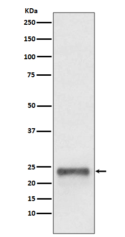
Western blot(SDS PAGE) analysis of extracts from HepG2 cell lysate.Using Ubiquitin D Rabbit mAb [A5S1]at dilution of 1:1000 incubated at 4℃ over night.
Product information
Protein names :FAT10; UBD-3; GABBR1; UBD; Ubiquitin D;
UniProtID :O15205
MASS(da) :18,473
MW(kDa) :23kDa
Form :Liquid
Purification :Affinity-chromatography
Host :Rabbit
Isotype : IgG
sensitivity :Endogenous
Reactivity :Human Mouse
- ApplicationDilution
- 免疫印迹(WB)1:1000-2000
- 免疫组化(IHC)1:100
- 免疫荧光(ICC/IF)1:100
- The optimal dilutions should be determined by the end user
Specificity :Antibody is produced by immunizing animals with A synthesized peptide derived from human Ubiquitin D
Storage :Antibody store in 10 mM PBS, 0.5mg/ml BSA, 50% glycerol. Shipped at 4°C. Store at-20°C or -80°C. Products are valid for one natural year of receipt.Avoid repeated freeze / thaw cycles.
WB Positive detected :HepG2 cell lysate.
Function : Ubiquitin-like protein modifier which can be covalently attached to target protein and subsequently leads to their degradation by the 26S proteasome, in a NUB1-dependent manner. Probably functions as a survival factor. Conjugation ability activated by UBA6. Promotes the expression of the proteasome subunit beta type-9 (PSMB9/LMP2). Regulates TNF-alpha-induced and LPS-mediated activation of the central mediator of innate immunity NF-kappa-B by promoting TNF-alpha-mediated proteasomal degradation of ubiquitinated-I-kappa-B-alpha. Required for TNF-alpha-induced p65 nuclear translocation in renal tubular epithelial cells (RTECs). May be involved in dendritic cell (DC) maturation, the process by which immature dendritic cells differentiate into fully competent antigen-presenting cells that initiate T-cell responses. Mediates mitotic non-disjunction and chromosome instability, in long-term in vitro culture and cancers, by abbreviating mitotic phase and impairing the kinetochore localization of MAD2L1 during the prometaphase stage of the cell cycle. May be involved in the formation of aggresomes when proteasome is saturated or impaired. Mediates apoptosis in a caspase-dependent manner, especially in renal epithelium and tubular cells during renal diseases such as polycystic kidney disease and Human immunodeficiency virus (HIV)-associated nephropathy (HIVAN)..
Tissue specificity :Constitutively expressed in mature dendritic cells and B-cells. Mostly expressed in the reticuloendothelial system (e.g. thymus, spleen), the gastrointestinal system, kidney, lung and prostate gland..
Subcellular locationi :Nucleus. Cytoplasm.
IMPORTANT: For western blots, incubate membrane with diluted primary antibody in 1% w/v BSA, 1X TBST at 4°C overnight.
-
About
-
Product
-
Law
-
Literature
-

WeChat scan code follow us