
NG2 Rabbit mAb [G3Bs]Cat NO.: A26915
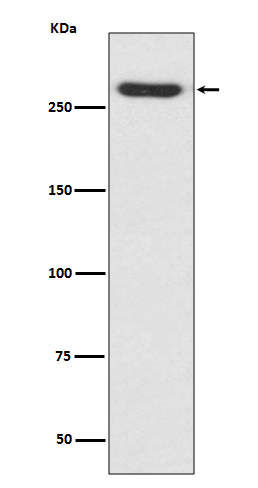
Western blot(SDS PAGE) analysis of extracts from A375 cell lysate.Using NG2 Rabbit mAb [G3Bs]at dilution of 1:1000 incubated at 4℃ over night.
Product information
Protein names :NG2; MCSP; MCSPG; MSK16; HMW-MAA; MEL-CSPG;
UniProtID :Q6UVK1
MASS(da) :250,537
MW(kDa) :251kDa
Form :Liquid
Purification :Affinity-chromatography
Host :Rabbit
Isotype : IgG
sensitivity :Endogenous
Reactivity :Human
- ApplicationDilution
- 免疫印迹(WB)1:1000-2000
- 免疫组化(IHC)1:100
- The optimal dilutions should be determined by the end user
Specificity :Antibody is produced by immunizing animals with A synthesized peptide derived from human NG2
Storage :Antibody store in 10 mM PBS, 0.5mg/ml BSA, 50% glycerol. Shipped at 4°C. Store at-20°C or -80°C. Products are valid for one natural year of receipt.Avoid repeated freeze / thaw cycles.
WB Positive detected :A375 cell lysate.
Function : Proteoglycan playing a role in cell proliferation and migration which stimulates endothelial cells motility during microvascular morphogenesis. May also inhibit neurite outgrowth and growth cone collapse during axon regeneration. Cell surface receptor for collagen alpha 2(VI) which may confer cells ability to migrate on that substrate. Binds through its extracellular N-terminus growth factors, extracellular matrix proteases modulating their activity. May regulate MPP16-dependent degradation and invasion of type I collagen participating in melanoma cells invasion properties. May modulate the plasminogen system by enhancing plasminogen activation and inhibiting angiostatin. Functions also as a signal transducing protein by binding through its cytoplasmic C-terminus scaffolding and signaling proteins. May promote retraction fiber formation and cell polarization through Rho GTPase activation. May stimulate alpha-4, beta-1 integrin-mediated adhesion and spreading by recruiting and activating a signaling cascade through CDC42, ACK1 and BCAR1. May activate FAK and ERK1/ERK2 signaling cascades..
Tissue specificity :Detected only in malignant melanoma cells..
Subcellular locationi :Cell membrane,Single-pass type I membrane protein,Extracellular side. Apical cell membrane,Single-pass type I membrane protein,Extracellular side. Cell projection, lamellipodium membrane,Single-pass type I membrane protein,Extracellular side. Cell surface.
IMPORTANT: For western blots, incubate membrane with diluted primary antibody in 1% w/v BSA, 1X TBST at 4°C overnight.
-
About
-
Product
-
Law
-
Literature
-

WeChat scan code follow us