
Podoplanin Rabbit mAb [L9v1]Cat NO.: A78021
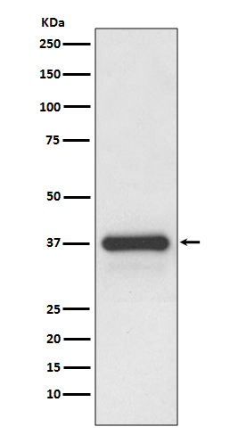
Western blot(SDS PAGE) analysis of extracts from human placenta lysate.Using Podoplanin Rabbit mAb [L9v1]at dilution of 1:1000 incubated at 4℃ over night.
Product information
Protein names :Aggrus; Glycoprotein 36 KD; GP36; GP38; GP40; HT1A1; hT1alpha1; hT1alpha2; OTS8; PA2.26; Pdpn; Podoplanin; T1 alpha; TI1A; TIA2;
UniProtID :Q86YL7
MASS(da) :16,698
MW(kDa) :36kDa
Form :Liquid
Purification :Affinity-chromatography
Host :Rabbit
Isotype : IgG
sensitivity :Endogenous
Reactivity :Human,Mouse,Rat
- ApplicationDilution
- 免疫印迹(WB)1:1000-2000
- The optimal dilutions should be determined by the end user
Specificity :Antibody is produced by immunizing animals with A synthesized peptide derived from human Podoplanin
Storage :Antibody store in 10 mM PBS, 0.5mg/ml BSA, 50% glycerol. Shipped at 4°C. Store at-20°C or -80°C. Products are valid for one natural year of receipt.Avoid repeated freeze / thaw cycles.
WB Positive detected :human placenta lysate.
Function : Mediates effects on cell migration and adhesion through its different partners. During development plays a role in blood and lymphatic vessels separation by binding CLEC1B, triggering CLEC1B activation in platelets and leading to platelet activation and/or aggregation (PubMed:14522983, PubMed:15231832, PubMed:17616532, PubMed:18215137, PubMed:17222411). Interaction with CD9, on the contrary, attenuates platelet aggregation induced by PDPN (PubMed:18541721). Through MSN or EZR interaction promotes epithelial-mesenchymal transition (EMT) leading to ERZ phosphorylation and triggering RHOA activation leading to cell migration increase and invasiveness (PubMed:17046996, PubMed:21376833). Interaction with CD44 promotes directional cell migration in epithelial and tumor cells (PubMed:20962267). In lymph nodes (LNs), controls fibroblastic reticular cells (FRCs) adhesion to the extracellular matrix (ECM) and contraction of the actomyosin by maintaining ERM proteins (EZR,MSN and RDX) and MYL9 activation through association with unknown transmembrane proteins. Engagement of CLEC1B by PDPN promotes FRCs relaxation by blocking lateral membrane interactions leading to reduction of ERM proteins (EZR,MSN and RDX) and MYL9 activation (By similarity). Through binding with LGALS8 may participate in connection of the lymphatic endothelium to the surrounding extracellular matrix (PubMed:19268462). In keratinocytes, induces changes in cell morphology showing an elongated shape, numerous membrane protrusions, major reorganization of the actin cytoskeleton, increased motility and decreased cell adhesion (PubMed:15515019). Controls invadopodia stability and maturation leading to efficient degradation of the extracellular matrix (ECM) in tumor cells through modulation of RHOC activity in order to activate ROCK1/ROCK2 and LIMK1/LIMK2 and inactivation of CFL1 (PubMed:25486435). Required for normal lung cell proliferation and alveolus formation at birth (By similarity). Does not function as a water channel or as a regulator of aquaporin-type water channels (PubMed:9651190). Does not have any effect on folic acid or amino acid transport (By similarity)..
Tissue specificity :Highly expressed in placenta, lung, skeletal muscle and brain. Weakly expressed in brain, kidney and liver. In placenta, expressed on the apical plasma membrane of endothelium. In lung, expressed in alveolar epithelium. Up-regulated in colorectal tumors and expressed in 25% of early oral squamous cell carcinomas..
Subcellular locationi :[Podoplanin]: Membrane,Single-pass type I membrane protein. Cell projection, lamellipodium membrane,Single-pass type I membrane protein. Cell projection, filopodium membrane,Single-pass type I membrane protein. Cell projection, microvillus membrane,Single-pass type I membrane protein. Cell projection, ruffle membrane,Single-pass type I membrane protein. Membrane raft. Apical cell membrane. Basolateral cell membrane. Cell projection, invadopodium.
IMPORTANT: For western blots, incubate membrane with diluted primary antibody in 1% w/v BSA, 1X TBST at 4°C overnight.
-
About
-
Product
-
Law
-
Literature
-

WeChat scan code follow us