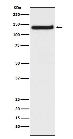
TLR9 Rabbit mAb [f717]Cat NO.: A40222
Western blot(SDS PAGE) analysis of extracts from Raji cell lysate.Using TLR9 Rabbit mAb [f717]at dilution of 1:1000 incubated at 4℃ over night.
Product information
Protein names :CD289; TLR9; Toll like receptor 9; Toll like receptor 9 isoform A precursor; Toll like receptor 9 isoform B;
UniProtID :Q9NR96
MASS(da) :115,860
MW(kDa) :130kDa
Form :Liquid
Purification :Affinity-chromatography
Host :Rabbit
Isotype : IgG
sensitivity :Endogenous
Reactivity :Human
- ApplicationDilution
- 免疫印迹(WB)1:1000-2000
- The optimal dilutions should be determined by the end user
Specificity :Antibody is produced by immunizing animals with A synthesized peptide derived from human TLR9
Storage :Antibody store in 10 mM PBS, 0.5mg/ml BSA, 50% glycerol. Shipped at 4°C. Store at-20°C or -80°C. Products are valid for one natural year of receipt.Avoid repeated freeze / thaw cycles.
WB Positive detected :Raji cell lysate.
Function : Key component of innate and adaptive immunity. TLRs (Toll-like receptors) control host immune response against pathogens through recognition of molecular patterns specific to microorganisms. TLR9 is a nucleotide-sensing TLR which is activated by unmethylated cytidine-phosphate-guanosine (CpG) dinucleotides. Acts via MYD88 and TRAF6, leading to NF-kappa-B activation, cytokine secretion and the inflammatory response (PubMed:11564765, PubMed:17932028). Controls lymphocyte response to Helicobacter infection (By similarity). Upon CpG stimulation, induces B-cell proliferation, activation, survival and antibody production (PubMed:23857366)..
Tissue specificity :Highly expressed in spleen, lymph node, tonsil and peripheral blood leukocytes, especially in plasmacytoid pre-dendritic cells. Levels are much lower in monocytes and CD11c+ immature dendritic cells. Also detected in lung and liver.
Subcellular locationi :Endoplasmic reticulum membrane,Single-pass type I membrane protein. Endosome. Lysosome. Cytoplasmic vesicle, phagosome.
IMPORTANT: For western blots, incubate membrane with diluted primary antibody in 1% w/v BSA, 1X TBST at 4°C overnight.


