BST2 Rabbit mAb [8Z2I]Cat NO.: A55379
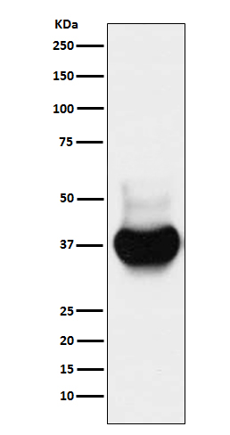
Western blot(SDS PAGE) analysis of extracts from HeLa cell lysate.Using BST2 Rabbit mAb [8Z2I]at dilution of 1:1000 incubated at 4℃ over night.
Product information
Protein names :Bone marrow stromal antigen 2; BST2; CD317; HM1.24 antigen; NPC A 7; Tetherin;
UniProtID :Q10589
MASS(da) :19,769
MW(kDa) :28-40kDa
Form :Liquid
Purification :Affinity-chromatography
Host :Rabbit
Isotype : IgG
sensitivity :Endogenous
Reactivity :Human
- ApplicationDilution
- 免疫印迹(WB)1:1000-2000
- 免疫组化(IHC)1:100
- The optimal dilutions should be determined by the end user
Specificity :Antibody is produced by immunizing animals with A synthesized peptide derived from human BST2
Storage :Antibody store in 10 mM PBS, 0.5mg/ml BSA, 50% glycerol. Shipped at 4°C. Store at-20°C or -80°C. Products are valid for one natural year of receipt.Avoid repeated freeze / thaw cycles.
WB Positive detected :HeLa cell lysate.
Function : IFN-induced antiviral host restriction factor which efficiently blocks the release of diverse mammalian enveloped viruses by directly tethering nascent virions to the membranes of infected cells. Acts as a direct physical tether, holding virions to the cell membrane and linking virions to each other. The tethered virions can be internalized by endocytosis and subsequently degraded or they can remain on the cell surface. In either case, their spread as cell-free virions is restricted (PubMed:22520941, PubMed:21529378, PubMed:20940320, PubMed:20419159, PubMed:20399176, PubMed:19879838, PubMed:19036818, PubMed:18342597, PubMed:18200009). Its target viruses belong to diverse families, including retroviridae: human immunodeficiency virus type 1 (HIV-1), human immunodeficiency virus type 2 (HIV-2), simian immunodeficiency viruses (SIVs), equine infectious anemia virus (EIAV), feline immunodeficiency virus (FIV), prototype foamy virus (PFV), Mason-Pfizer monkey virus (MPMV), human T-cell leukemia virus type 1 (HTLV-1), Rous sarcoma virus (RSV) and murine leukemia virus (MLV), flavivirideae: hepatitis C virus (HCV), filoviridae: ebola virus (EBOV) and marburg virus (MARV), arenaviridae: lassa virus (LASV) and machupo virus (MACV), herpesviridae: kaposis sarcoma-associated herpesvirus (KSHV), rhabdoviridae: vesicular stomatitis virus (VSV), orthomyxoviridae: influenza A virus, paramyxoviridae: nipah virus, and coronaviridae: SARS-CoV (PubMed:22520941, PubMed:21621240, PubMed:21529378, PubMed:20943977, PubMed:20686043, PubMed:20419159, PubMed:20399176, PubMed:19879838, PubMed:19179289, PubMed:18342597, PubMed:18200009, PubMed:26378163, PubMed:31199522). Can inhibit cell surface proteolytic activity of MMP14 causing decreased activation of MMP15 which results in inhibition of cell growth and migration (PubMed:22065321). Can stimulate signaling by LILRA4/ILT7 and consequently provide negative feedback to the production of IFN by plasmacytoid dendritic cells in response to viral infection (PubMed:19564354, PubMed:26172439). Plays a role in the organization of the subapical actin cytoskeleton in polarized epithelial cells. Isoform 1 and isoform 2 are both effective viral restriction factors but have differing antiviral and signaling activities (PubMed:23028328, PubMed:26172439). Isoform 2 is resistant to HIV-1 Vpu-mediated degradation and restricts HIV-1 viral budding in the presence of Vpu (PubMed:23028328, PubMed:26172439). Isoform 1 acts as an activator of NF-kappa-B and this activity is inhibited by isoform 2 (PubMed:23028328)..
Tissue specificity :Predominantly expressed in liver, lung, heart and placenta. Lower levels in pancreas, kidney, skeletal muscle and brain. Overexpressed in multiple myeloma cells. Highly expressed during B-cell development, from pro-B precursors to plasma cells. Highly expressed on T-cells, monocytes, NK cells and dendritic cells (at protein level)..
Subcellular locationi :Golgi apparatus, trans-Golgi network. Cell membrane,Single-pass type II membrane protein. Cell membrane,Lipid-anchor, GPI-anchor. Membrane raft. Cytoplasm. Apical cell membrane.
IMPORTANT: For western blots, incubate membrane with diluted primary antibody in 1% w/v BSA, 1X TBST at 4°C overnight.


