
ULK3 Rabbit mAb [8xAS]Cat NO.: A32652
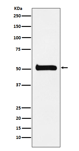
Western blot(SDS PAGE) analysis of extracts from 293T cell lysate.Using ULK3 Rabbit mAb [8xAS]at dilution of 1:1000 incubated at 4℃ over night.
Product information
Protein names :Serine/threonine-protein kinase ULK3; Ulk3; unc 51 like kinase 3 (C. elegans); Unc-51-like kinase 3;
UniProtID :Q6PHR2
MASS(da) :53,444
MW(kDa) :53kDa
Form :Liquid
Purification :Affinity-chromatography
Host :Rabbit
Isotype : IgG
sensitivity :Endogenous
Reactivity :Human,Mouse,Rat
- ApplicationDilution
- 免疫印迹(WB)1:1000-2000
- 免疫组化(IHC)1:100
- The optimal dilutions should be determined by the end user
Specificity :Antibody is produced by immunizing animals with A synthesized peptide derived from human ULK3
Storage :Antibody store in 10 mM PBS, 0.5mg/ml BSA, 50% glycerol. Shipped at 4°C. Store at-20°C or -80°C. Products are valid for one natural year of receipt.Avoid repeated freeze / thaw cycles.
WB Positive detected :293T cell lysate.
Function : Serine/threonine protein kinase that acts as a regulator of Sonic hedgehog (SHH) signaling and autophagy. Acts as a negative regulator of SHH signaling in the absence of SHH ligand: interacts with SUFU, thereby inactivating the protein kinase activity and preventing phosphorylation of GLI proteins (GLI1, GLI2 and/or GLI3). Positively regulates SHH signaling in the presence of SHH: dissociates from SUFU, autophosphorylates and mediates phosphorylation of GLI2, activating it and promoting its nuclear translocation. Phosphorylates in vitro GLI2, as well as GLI1 and GLI3, although less efficiently. Also acts as a regulator of autophagy: following cellular senescence, able to induce autophagy..
Tissue specificity :Widely expressed. Highest levels observed in fetal brain. In adult tissues, high levels in brain, liver and kidney, moderate levels in testis and adrenal gland and low levels in heart, lung, stomach, thymus, prostate and placenta. In the brain, highest expression in the hippocampus, high levels also detected in the cerebellum, olfactory bulb and optic nerve. In the central nervous system, lowest levels in the spinal cord..
Subcellular locationi :Cytoplasm.
IMPORTANT: For western blots, incubate membrane with diluted primary antibody in 1% w/v BSA, 1X TBST at 4°C overnight.
-
About
-
Product
-
Law
-
Literature
-

WeChat scan code follow us