
PAK2 Rabbit mAb [jIS5]Cat NO.: A55075
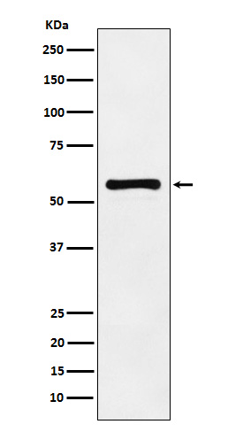
Western blot(SDS PAGE) analysis of extracts from HeLa cell lysate.Using PAK2 Rabbit mAb [jIS5]at dilution of 1:1000 incubated at 4℃ over night.
Product information
Protein names :CB422; Gamma PAK; hPAK65; p27; p34; p58; p65PAK; PAK-2p34; Pak2; PAK65; PAKgamma; S6 H4 kinase;
UniProtID :Q13177
MASS(da) :58,043
MW(kDa) :61kDa
Form :Liquid
Purification :Affinity-chromatography
Host :Rabbit
Isotype : IgG
sensitivity :Endogenous
Reactivity :Human,Mouse,Rat
- ApplicationDilution
- 免疫印迹(WB)1:1000-2000
- 免疫组化(IHC)1:100
- 免疫荧光(ICC/IF)1:100
- The optimal dilutions should be determined by the end user
Specificity :Antibody is produced by immunizing animals with A synthesized peptide derived from human PAK2
Storage :Antibody store in 10 mM PBS, 0.5mg/ml BSA, 50% glycerol. Shipped at 4°C. Store at-20°C or -80°C. Products are valid for one natural year of receipt.Avoid repeated freeze / thaw cycles.
WB Positive detected :HeLa cell lysate.
Function : Serine/threonine protein kinase that plays a role in a variety of different signaling pathways including cytoskeleton regulation, cell motility, cell cycle progression, apoptosis or proliferation. Acts as downstream effector of the small GTPases CDC42 and RAC1. Activation by the binding of active CDC42 and RAC1 results in a conformational change and a subsequent autophosphorylation on several serine and/or threonine residues. Full-length PAK2 stimulates cell survival and cell growth. Phosphorylates MAPK4 and MAPK6 and activates the downstream target MAPKAPK5, a regulator of F-actin polymerization and cell migration. Phosphorylates JUN and plays an important role in EGF-induced cell proliferation. Phosphorylates many other substrates including histone H4 to promote assembly of H3.3 and H4 into nucleosomes, BAD, ribosomal protein S6, or MBP. Additionally, associates with ARHGEF7 and GIT1 to perform kinase-independent functions such as spindle orientation control during mitosis. On the other hand, apoptotic stimuli such as DNA damage lead to caspase-mediated cleavage of PAK2, generating PAK-2p34, an active p34 fragment that translocates to the nucleus and promotes cellular apoptosis involving the JNK signaling pathway. Caspase-activated PAK2 phosphorylates MKNK1 and reduces cellular translation..
Tissue specificity :Ubiquitously expressed. Higher levels seen in skeletal muscle, ovary, thymus and spleen.
Subcellular locationi :[Serine/threonine-protein kinase PAK 2]: Cytoplasm.
IMPORTANT: For western blots, incubate membrane with diluted primary antibody in 1% w/v BSA, 1X TBST at 4°C overnight.
-
About
-
Product
-
Law
-
Literature
-

WeChat scan code follow us