SMURF 2 Rabbit mAb [O1XZ]Cat NO.: A79823
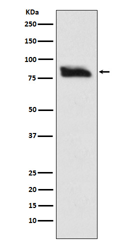
Western blot(SDS PAGE) analysis of extracts from SH-SY-5Y cell lysate.Using SMURF 2 Rabbit mAb [O1XZ]at dilution of 1:1000 incubated at 4℃ over night.
Product information
Protein names :hSMURF2; SMUF2_HUMAN; Smurf2;
UniProtID :Q9HAU4
MASS(da) :86,196
MW(kDa) :86kDa
Form :Liquid
Purification :Affinity-chromatography
Host :Rabbit
Isotype : IgG
sensitivity :Endogenous
Reactivity :Human,Mouse,Rat
- ApplicationDilution
- 免疫印迹(WB)1:1000-2000
- The optimal dilutions should be determined by the end user
Specificity :Antibody is produced by immunizing animals with A synthesized peptide derived from human SMURF 2
Storage :Antibody store in 10 mM PBS, 0.5mg/ml BSA, 50% glycerol. Shipped at 4°C. Store at-20°C or -80°C. Products are valid for one natural year of receipt.Avoid repeated freeze / thaw cycles.
WB Positive detected :SH-SY-5Y cell lysate.
Function : E3 ubiquitin-protein ligase which accepts ubiquitin from an E2 ubiquitin-conjugating enzyme in the form of a thioester and then directly transfers the ubiquitin to targeted substrates (PubMed:11016919). Interacts with SMAD7 to trigger SMAD7-mediated transforming growth factor beta/TGF-beta receptor ubiquitin-dependent degradation, thereby down-regulating TGF-beta signaling (PubMed:11163210, PubMed:12717440). In addition, interaction with SMAD7 activates autocatalytic degradation, which is prevented by interaction with AIMP1 (PubMed:18448069). Also forms a stable complex with TGF-beta receptor-mediated phosphorylated SMAD1, SMAD2 and SMAD3, and targets SMAD1 and SMAD2 for ubiquitination and proteasome-mediated degradation (PubMed:11016919, PubMed:11158580, PubMed:11389444). SMAD2 may recruit substrates, such as SNON, for ubiquitin-dependent degradation (PubMed:11389444). Negatively regulates TGFB1-induced epithelial-mesenchymal transition and myofibroblast differentiation (PubMed:30696809).., (Microbial infection) In case of filoviruses Ebola/EBOV and Marburg/MARV infection, the complex formed by viral matrix protein VP40 and SMURF2 facilitates virus budding..
Tissue specificity :Widely expressed.
Subcellular locationi :Nucleus. Cytoplasm. Cell membrane. Membrane raft.
IMPORTANT: For western blots, incubate membrane with diluted primary antibody in 1% w/v BSA, 1X TBST at 4°C overnight.


