
IMP3 Rabbit mAb [HdE8]Cat NO.: A95572
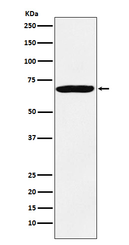
Western blot(SDS PAGE) analysis of extracts from HeLa cell lysate.Using IMP3 Rabbit mAb [HdE8]at dilution of 1:1000 incubated at 4℃ over night.
Product information
Protein names :CT98; hKOC; IGF2BP3; IMP 3; KOC1; VICKZ 3;
UniProtID :O00425
MASS(da) :63,705
MW(kDa) :69kDa
Form :Liquid
Purification :Affinity-chromatography
Host :Rabbit
Isotype : IgG
sensitivity :Endogenous
Reactivity :Human,Mouse,Rat
- ApplicationDilution
- 免疫印迹(WB)1:1000-2000
- 免疫荧光(ICC/IF)1:100
- The optimal dilutions should be determined by the end user
Specificity :Antibody is produced by immunizing animals with A synthesized peptide derived from human IMP3
Storage :Antibody store in 10 mM PBS, 0.5mg/ml BSA, 50% glycerol. Shipped at 4°C. Store at-20°C or -80°C. Products are valid for one natural year of receipt.Avoid repeated freeze / thaw cycles.
WB Positive detected :HeLa cell lysate.
Function : RNA-binding factor that may recruit target transcripts to cytoplasmic protein-RNA complexes (mRNPs). This transcript 'caging' into mRNPs allows mRNA transport and transient storage. It also modulates the rate and location at which target transcripts encounter the translational apparatus and shields them from endonuclease attacks or microRNA-mediated degradation. Preferentially binds to N6-methyladenosine (m6A)-containing mRNAs and increases their stability (PubMed:29476152). Binds to the 3'-UTR of CD44 mRNA and stabilizes it, hence promotes cell adhesion and invadopodia formation in cancer cells. Binds to beta-actin/ACTB and MYC transcripts. Increases MYC mRNA stability by binding to the coding region instability determinant (CRD) and binding is enhanced by m6A-modification of the CRD (PubMed:29476152). Binds to the 5'-UTR of the insulin-like growth factor 2 (IGF2) mRNAs..
Tissue specificity :Expressed in fetal liver, fetal lung, fetal kidney, fetal thymus, fetal placenta, fetal follicles of ovary and gonocytes of testis, growing oocytes, spermatogonia and semen (at protein level). Expressed in cervix adenocarcinoma, in testicular, pancreatic and renal-cell carcinomas (at protein level). Expressed ubiquitously during fetal development at 8 and 14 weeks of gestation. Expressed in ovary, testis, brain, placenta, pancreatic cancer tissues and pancreatic cancer cell lines..
Subcellular locationi :Nucleus. Cytoplasm. Cytoplasm, P-body. Cytoplasm, Stress granule.
IMPORTANT: For western blots, incubate membrane with diluted primary antibody in 1% w/v BSA, 1X TBST at 4°C overnight.
-
About
-
Product
-
Law
-
Literature
-

WeChat scan code follow us