Histone H1.2 Rabbit mAb [lFb7]Cat NO.: A31401
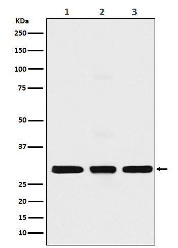
Western blot(SDS PAGE) analysis of extracts from (1) MCF7 cell lysate(2) NIH/3T3 cell lysate; (3) C6 cell lysate.Using Histone H1.2 Rabbit mAb [lFb7]at dilution of 1:1000 incubated at 4℃ over night.
Product information
Protein names :H1.a; H1F2; H1s-1; HIST1H1C; Histone H1.2; Histone H1c; Histone H1d; Histone H1s-1;
UniProtID :P16403
MASS(da) :21,365
MW(kDa) :29kDa
Form :Liquid
Purification :Affinity-chromatography
Host :Rabbit
Isotype : IgG
sensitivity :Endogenous
Reactivity :Human,Mouse,Rat
- ApplicationDilution
- 免疫印迹(WB)1:1000-2000
- 免疫组化(IHC)1:100
- 免疫荧光(ICC/IF)1:100
- The optimal dilutions should be determined by the end user
Specificity :Antibody is produced by immunizing animals with A synthesized peptide derived from human Histone H1.2
Storage :Antibody store in 10 mM PBS, 0.5mg/ml BSA, 50% glycerol. Shipped at 4°C. Store at-20°C or -80°C. Products are valid for one natural year of receipt.Avoid repeated freeze / thaw cycles.
WB Positive detected :(1) MCF7 cell lysate(2) NIH/3T3 cell lysate; (3) C6 cell lysate.
Function : Histone H1 protein binds to linker DNA between nucleosomes forming the macromolecular structure known as the chromatin fiber. Histones H1 are necessary for the condensation of nucleosome chains into higher-order structured fibers. Acts also as a regulator of individual gene transcription through chromatin remodeling, nucleosome spacing and DNA methylation (By similarity)..
Subcellular locationi :Nucleus. Chromosome.
IMPORTANT: For western blots, incubate membrane with diluted primary antibody in 1% w/v BSA, 1X TBST at 4°C overnight.


