
MIB1 / DIP1 Rabbit mAb [D62V]Cat NO.: A72941
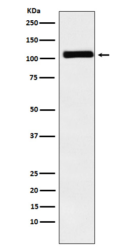
Western blot(SDS PAGE) analysis of extracts from Caco2 cell lysate.Using MIB1 / DIP1 Rabbit mAb [D62V]at dilution of 1:1000 incubated at 4℃ over night.
Product information
Protein names :Dip1; LVNC7; MIB; mib1; ZZANK2; ZZZ6;
UniProtID :Q86YT6
MASS(da) :110,136
MW(kDa) :110kDa
Form :Liquid
Purification :Affinity-chromatography
Host :Rabbit
Isotype : IgG
sensitivity :Endogenous
Reactivity :Human Mouse
- ApplicationDilution
- 免疫印迹(WB)1:1000-2000
- 免疫组化(IHC)1:100
- 免疫荧光(ICC/IF)1:100
- The optimal dilutions should be determined by the end user
Specificity :Antibody is produced by immunizing animals with A synthesized peptide derived from human MIB1 / DIP1
Storage :Antibody store in 10 mM PBS, 0.5mg/ml BSA, 50% glycerol. Shipped at 4°C. Store at-20°C or -80°C. Products are valid for one natural year of receipt.Avoid repeated freeze / thaw cycles.
WB Positive detected :Caco2 cell lysate.
Function : E3 ubiquitin-protein ligase that mediates ubiquitination of Delta receptors, which act as ligands of Notch proteins. Positively regulates the Delta-mediated Notch signaling by ubiquitinating the intracellular domain of Delta, leading to endocytosis of Delta receptors. Probably mediates ubiquitination and subsequent proteasomal degradation of DAPK1, thereby antagonizing anti-apoptotic effects of DAPK1 to promote TNF-induced apoptosis (By similarity). Involved in ubiquitination of centriolar satellite CEP131, CEP290 and PCM1 proteins and hence inhibits primary cilium formation in proliferating cells. Mediates 'Lys-63'-linked polyubiquitination of TBK1, which probably participates in kinase activation..
Tissue specificity :Widely expressed at low level. Expressed at higher level in spinal cord, ovary, whole brain, and all specific brain regions examined..
Subcellular locationi :Cytoplasm. Cytoplasm, cytoskeleton, microtubule organizing center, centrosome, centriolar satellite. Cell membrane.
IMPORTANT: For western blots, incubate membrane with diluted primary antibody in 1% w/v BSA, 1X TBST at 4°C overnight.
-
About
-
Product
-
Law
-
Literature
-

WeChat scan code follow us