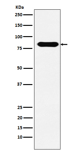
CD98 Rabbit mAb [JJ1v]Cat NO.: A66413
Western blot(SDS PAGE) analysis of extracts from HepG2 cell lysate.Using CD98 Rabbit mAb [JJ1v]at dilution of 1:1000 incubated at 4℃ over night.
Product information
Protein names :4T2HC; CD98; CD98HC; MDU1; NACAE; Slc3a2;
UniProtID :P08195
MASS(da) :67,994
MW(kDa) :80kDa
Form :Liquid
Purification :Affinity-chromatography
Host :Rabbit
Isotype : IgG
sensitivity :Endogenous
Reactivity :Human
- ApplicationDilution
- 免疫印迹(WB)1:1000-2000
- 免疫组化(IHC)1:100
- The optimal dilutions should be determined by the end user
Specificity :Antibody is produced by immunizing animals with A synthesized peptide derived from human CD98
Storage :Antibody store in 10 mM PBS, 0.5mg/ml BSA, 50% glycerol. Shipped at 4°C. Store at-20°C or -80°C. Products are valid for one natural year of receipt.Avoid repeated freeze / thaw cycles.
WB Positive detected :HepG2 cell lysate.
Function : Component of several heterodimeric complexes involved in amino acid transport (PubMed:11557028, PubMed:9829974, PubMed:9751058, PubMed:10391915, PubMed:10574970, PubMed:11311135, PubMed:30341327). The precise substrate specificity depends on the other subunit in the heterodimer (PubMed:9829974, PubMed:9751058, PubMed:10391915, PubMed:10574970, PubMed:30867591, PubMed:10903140). The complexes function as amino acid exchangers (PubMed:11557028, PubMed:10903140, PubMed:12117417, PubMed:12225859, PubMed:30867591). The homodimer functions as sodium-independent, high-affinity transporter that mediates uptake of large neutral amino acids such as phenylalanine, tyrosine, L-DOPA, leucine, histidine, methionine and tryptophan (PubMed:9751058, PubMed:11557028, PubMed:11311135, PubMed:11564694, PubMed:12117417, PubMed:12225859, PubMed:25998567, PubMed:30867591). The heterodimer formed by SLC3A2 and SLC7A6 or SLC3A2 and SLC7A7 mediates the uptake of dibasic amino acids (PubMed:9829974, PubMed:10903140). The heterodimer with SLC7A5/LAT1 mediates the transport of thyroid hormones triiodothyronine (T3) and thyroxine (T4) across the cell membrane (PubMed:11564694, PubMed:12225859). The heterodimer with SLC7A5/LAT1 is involved in the uptake of toxic methylmercury (MeHg) when administered as the L-cysteine or D,L-homocysteine complexes (PubMed:12117417). The heterodimer with SLC7A5/LAT1 is involved in the uptake of leucine (PubMed:25998567, PubMed:30341327). When associated with LAPTM4B, the heterodimer with SLC7A5/LAT1 is recruited to lysosomes to promote leucine uptake into these organelles, and thereby mediates mTORC1 activation (PubMed:25998567). The heterodimer with SLC7A5/LAT1 may play a role in the transport of L-DOPA across the blood-brain barrier (By similarity). The heterodimer formed by SLC3A2 and SLC7A5/LAT1 or SLC3A2 and SLC7A8/LAT2 is involved in the cellular activity of small molecular weight nitrosothiols, via the stereoselective transport of L-nitrosocysteine (L-CNSO) across the transmembrane (PubMed:15769744). Together with ICAM1, regulates the transport activity of SLC7A8/LAT2 in polarized intestinal cells by generating and delivering intracellular signals (PubMed:12716892). Required for targeting of SLC7A5/LAT1 and SLC7A8/LAT2 to the plasma membrane and for channel activity (PubMed:9751058, PubMed:11311135, PubMed:30867591). Plays a role in nitric oxide synthesis in human umbilical vein endothelial cells (HUVECs) via transport of L-arginine (PubMed:14603368). May mediate blood-to-retina L-leucine transport across the inner blood-retinal barrier (By similarity).., (Microbial infection) In case of hepatitis C virus/HCV infection, the complex formed by SLC3A2 and SLC7A5/LAT1 plays a role in HCV propagation by facilitating viral entry into host cell and increasing L-leucine uptake-mediated mTORC1 signaling activation, thereby contributing to HCV-mediated pathogenesis..
Tissue specificity :Expressed ubiquitously in all tissues tested with highest levels detected in kidney, placenta and testis and weakest level in thymus. During gestation, expression in the placenta was significantly stronger at full-term than at the mid-trimester stage. Expressed in HUVECS and at low levels in resting peripheral blood T-lymphocytes and quiescent fibroblasts. Also expressed in fetal liver and in the astrocytic process of primary astrocytic gliomas. Expressed in retinal endothelial cells and in the intestinal epithelial cell line C2BBe1..
Subcellular locationi :Apical cell membrane. Cell membrane,Single-pass type II membrane protein. Cell junction. Lysosome membrane. Melanosome.
IMPORTANT: For western blots, incubate membrane with diluted primary antibody in 1% w/v BSA, 1X TBST at 4°C overnight.


