
Plastin L Rabbit mAb [WDU7]Cat NO.: A82835
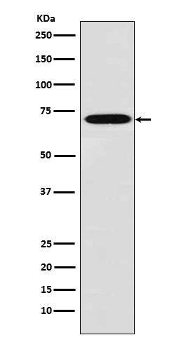
Western blot(SDS PAGE) analysis of extracts from THP1 cell lysate.Using Plastin L Rabbit mAb [WDU7]at dilution of 1:1000 incubated at 4℃ over night.
Product information
Protein names :CP64; L plastin; LC64P; LCP1; LPL; Lplastin; Plastin 2; PLS2;
UniProtID :P13796
MASS(da) :70,288
MW(kDa) :70kDa
Form :Liquid
Purification :Affinity-chromatography
Host :Rabbit
Isotype : IgG
sensitivity :Endogenous
Reactivity :Human,Mouse,Rat
- ApplicationDilution
- 免疫印迹(WB)1:1000-2000
- 免疫组化(IHC)1:100
- 免疫荧光(ICC/IF)1:100
- The optimal dilutions should be determined by the end user
Specificity :Antibody is produced by immunizing animals with A synthesized peptide derived from human Plastin L
Storage :Antibody store in 10 mM PBS, 0.5mg/ml BSA, 50% glycerol. Shipped at 4°C. Store at-20°C or -80°C. Products are valid for one natural year of receipt.Avoid repeated freeze / thaw cycles.
WB Positive detected :THP1 cell lysate.
Function : Actin-binding protein (PubMed:16636079, PubMed:17294403, PubMed:28493397). Plays a role in the activation of T-cells in response to costimulation through TCR/CD3 and CD2 or CD28 (PubMed:17294403). Modulates the cell surface expression of IL2RA/CD25 and CD69 (PubMed:17294403)..
Tissue specificity :Detected in intestinal microvilli, hair cell stereocilia, and fibroblast filopodia, in spleen and other lymph node-containing organs. Expressed in peripheral blood T-lymphocytes, neutrophils, monocytes, B-lymphocytes, and myeloid cells..
Subcellular locationi :Cytoplasm, cytoskeleton. Cell junction. Cell projection. Cell projection, ruffle membrane,Peripheral membrane protein,Cytoplasmic side.
IMPORTANT: For western blots, incubate membrane with diluted primary antibody in 1% w/v BSA, 1X TBST at 4°C overnight.
-
About
-
Product
-
Law
-
Literature
-

WeChat scan code follow us