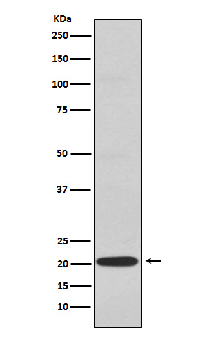
CD3G Rabbit mAb [X3HW]Cat NO.: A98219
Western blot(SDS PAGE) analysis of extracts from Jurkat cell lysate.Using CD3G Rabbit mAb [X3HW]at dilution of 1:1000 incubated at 4℃ over night.
Product information
Protein names :CD3 gamma; CD3g; T3G;
UniProtID :P09693
MASS(da) :20,469
MW(kDa) :20kDa
Form :Liquid
Purification :Affinity-chromatography
Host :Rabbit
Isotype : IgG
sensitivity :Endogenous
Reactivity :Human Mouse
- ApplicationDilution
- 免疫印迹(WB)1:1000-2000
- 免疫组化(IHC)1:100
- 免疫荧光(ICC/IF)1:100
- The optimal dilutions should be determined by the end user
Specificity :Antibody is produced by immunizing animals with A synthesized peptide derived from human CD3G
Storage :Antibody store in 10 mM PBS, 0.5mg/ml BSA, 50% glycerol. Shipped at 4°C. Store at-20°C or -80°C. Products are valid for one natural year of receipt.Avoid repeated freeze / thaw cycles.
WB Positive detected :Jurkat cell lysate.
Function : Part of the TCR-CD3 complex present on T-lymphocyte cell surface that plays an essential role in adaptive immune response. When antigen presenting cells (APCs) activate T-cell receptor (TCR), TCR-mediated signals are transmitted across the cell membrane by the CD3 chains CD3D, CD3E, CD3G and CD3Z. All CD3 chains contain immunoreceptor tyrosine-based activation motifs (ITAMs) in their cytoplasmic domain. Upon TCR engagement, these motifs become phosphorylated by Src family protein tyrosine kinases LCK and FYN, resulting in the activation of downstream signaling pathways (PubMed:2470098). In addition to this role of signal transduction in T-cell activation, CD3G plays an essential role in the dynamic regulation of TCR expression at the cell surface (PubMed:8187769). Indeed, constitutive TCR cycling is dependent on the di-leucine-based (diL) receptor-sorting motif present in CD3G..
Subcellular locationi :Cell membrane,Single-pass type I membrane protein.
IMPORTANT: For western blots, incubate membrane with diluted primary antibody in 1% w/v BSA, 1X TBST at 4°C overnight.


