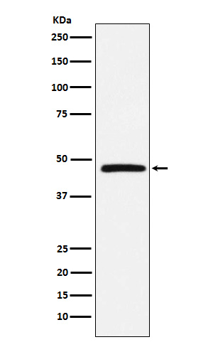
DARC Rabbit mAb [nf70]Cat NO.: A72753
Western blot(SDS PAGE) analysis of extracts from Human fetal liver lysate.Using DARC Rabbit mAb [nf70]at dilution of 1:1000 incubated at 4℃ over night.
Product information
Protein names :CCBP1; CD234; DARC; Dfy; FY; GPD; GpFy; WBCQ1;
UniProtID :Q16570
MASS(da) :35,553
MW(kDa) :35kDa
Form :Liquid
Purification :Affinity-chromatography
Host :Rabbit
Isotype : IgG
sensitivity :Endogenous
Reactivity :Human Mouse
- ApplicationDilution
- 免疫印迹(WB)1:1000-2000
- 免疫组化(IHC)1:100
- 免疫荧光(ICC/IF)1:100
- The optimal dilutions should be determined by the end user
Specificity :Antibody is produced by immunizing animals with A synthesized peptide derived from human DARC
Storage :Antibody store in 10 mM PBS, 0.5mg/ml BSA, 50% glycerol. Shipped at 4°C. Store at-20°C or -80°C. Products are valid for one natural year of receipt.Avoid repeated freeze / thaw cycles.
WB Positive detected :Human fetal liver lysate.
Function : Atypical chemokine receptor that controls chemokine levels and localization via high-affinity chemokine binding that is uncoupled from classic ligand-driven signal transduction cascades, resulting instead in chemokine sequestration, degradation, or transcytosis. Also known as interceptor (internalizing receptor) or chemokine-scavenging receptor or chemokine decoy receptor. Has a promiscuous chemokine-binding profile, interacting with inflammatory chemokines of both the CXC and the CC subfamilies but not with homeostatic chemokines. Acts as a receptor for chemokines including CCL2, CCL5, CCL7, CCL11, CCL13, CCL14, CCL17, CXCL5, CXCL6, IL8/CXCL8, CXCL11, GRO, RANTES, MCP-1, TARC and also for the malaria parasites P.vivax and P.knowlesi. May regulate chemokine bioavailability and, consequently, leukocyte recruitment through two distinct mechanisms: when expressed in endothelial cells, it sustains the abluminal to luminal transcytosis of tissue-derived chemokines and their subsequent presentation to circulating leukocytes,when expressed in erythrocytes, serves as blood reservoir of cognate chemokines but also as a chemokine sink, buffering potential surges in plasma chemokine levels.
Tissue specificity :Found in adult kidney, adult spleen, bone marrow and fetal liver. In particular, it is expressed along postcapillary venules throughout the body, except in the adult liver. Erythroid cells and postcapillary venule endothelium are the principle tissues expressing duffy. Fy(-A-B) individuals do not express duffy in the bone marrow, however they do, in postcapillary venule endothelium.
Subcellular locationi :Early endosome. Recycling endosome. Membrane,Multi-pass membrane protein.
IMPORTANT: For western blots, incubate membrane with diluted primary antibody in 1% w/v BSA, 1X TBST at 4°C overnight.


