
IL23 Rabbit mAb [F8a4]Cat NO.: A92423
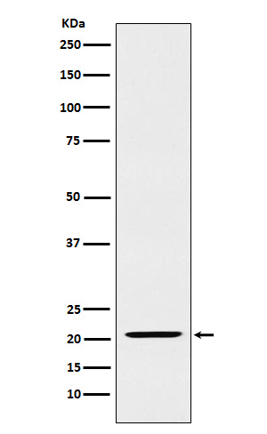
Western blot(SDS PAGE) analysis of extracts from Raji cell lysate.Using IL23 Rabbit mAb [F8a4]at dilution of 1:1000 incubated at 4℃ over night.
Product information
Protein names :IL 23A; IL 23p19; IL12B; IL23; Il23a; IL23P19; P19; SGRF;
UniProtID :Q9NPF7
MASS(da) :20,730
MW(kDa) :21kDa
Form :Liquid
Purification :Affinity-chromatography
Host :Rabbit
Isotype : IgG
sensitivity :Endogenous
Reactivity :Human
- ApplicationDilution
- 免疫印迹(WB)1:1000-2000
- The optimal dilutions should be determined by the end user
Specificity :Antibody is produced by immunizing animals with A synthesized peptide derived from human IL23
Storage :Antibody store in 10 mM PBS, 0.5mg/ml BSA, 50% glycerol. Shipped at 4°C. Store at-20°C or -80°C. Products are valid for one natural year of receipt.Avoid repeated freeze / thaw cycles.
WB Positive detected :Raji cell lysate.
Function : Associates with IL12B to form the pro-inflammatory cytokine IL-23 that plays different roles in innate and adaptive immunity (PubMed:11114383). Released by antigen-presenting cells such as dendritic cells or macrophages, binds to a heterodimeric receptor complex composed of IL12RB1 and IL23R to activate JAK2 and TYK2 which then phosphorylate the receptor to form a docking site leading to the phosphorylation of STAT3 and STAT4 (PubMed:32474165, PubMed:29287995, PubMed:33606986). This process leads to activation of several pathways including p38 MAPK or NF-kappa-B and promotes the production of pro-inflammatory cytokines such as interleukin-17A/IL17A (PubMed:12023369). In turn, participates in the early and effective intracellular bacterial clearance (PubMed:32474165). Promotes the expansion and survival of T-helper 17 cells, a CD4-positive helper T-cell subset that produces IL-17, as well as other IL-17-producing cells (PubMed:17676044)..
Tissue specificity :Secreted by activated dendritic and phagocytic cells and keratinocytes. Also expressed by dermal Langerhans cells (at protein level)..
Subcellular locationi :Secreted.
IMPORTANT: For western blots, incubate membrane with diluted primary antibody in 1% w/v BSA, 1X TBST at 4°C overnight.
-
About
-
Product
-
Law
-
Literature
-

WeChat scan code follow us