NEK2 Rabbit mAb [CFor]Cat NO.: A17041
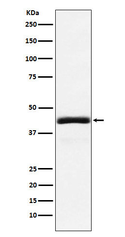
Western blot(SDS PAGE) analysis of extracts from 293T cell lysate.Using NEK2 Rabbit mAb [CFor]at dilution of 1:1000 incubated at 4℃ over night.
Product information
Protein names :HsPK21; NEK2; NEK2A; NLK1;
UniProtID :P51955
MASS(da) :51,763
MW(kDa) :48/44kDa
Form :Liquid
Purification :Affinity-chromatography
Host :Rabbit
Isotype : IgG
sensitivity :Endogenous
Reactivity :Human
- ApplicationDilution
- 免疫印迹(WB)1:1000-2000
- The optimal dilutions should be determined by the end user
Specificity :Antibody is produced by immunizing animals with A synthesized peptide derived from human NEK2
Storage :Antibody store in 10 mM PBS, 0.5mg/ml BSA, 50% glycerol. Shipped at 4°C. Store at-20°C or -80°C. Products are valid for one natural year of receipt.Avoid repeated freeze / thaw cycles.
WB Positive detected :293T cell lysate.
Function : Protein kinase which is involved in the control of centrosome separation and bipolar spindle formation in mitotic cells and chromatin condensation in meiotic cells. Regulates centrosome separation (essential for the formation of bipolar spindles and high-fidelity chromosome separation) by phosphorylating centrosomal proteins such as CROCC, CEP250 and NINL, resulting in their displacement from the centrosomes. Regulates kinetochore microtubule attachment stability in mitosis via phosphorylation of NDC80. Involved in regulation of mitotic checkpoint protein complex via phosphorylation of CDC20 and MAD2L1. Plays an active role in chromatin condensation during the first meiotic division through phosphorylation of HMGA2. Phosphorylates: PPP1CC,SGO1,NECAB3 and NPM1. Essential for localization of MAD2L1 to kinetochore and MAPK1 and NPM1 to the centrosome. Phosphorylates CEP68 and CNTLN directly or indirectly (PubMed:24554434). NEK2-mediated phosphorylation of CEP68 promotes CEP68 dissociation from the centrosome and its degradation at the onset of mitosis (PubMed:25704143). Involved in the regulation of centrosome disjunction (PubMed:26220856).., [Isoform 1]: Phosphorylates and activates NEK11 in G1/S-arrested cells.., [Isoform 2]: Not present in the nucleolus and, in contrast to isoform 1, does not phosphorylate and activate NEK11 in G1/S-arrested cells..
Tissue specificity :Isoform 1 and isoform 2 are expressed in peripheral blood T-cells and a wide variety of transformed cell types. Isoform 1 and isoform 4 are expressed in the testis. Up-regulated in various cancer cell lines, as well as primary breast tumors..
Subcellular locationi :[Isoform 1]: Nucleus. Nucleus, nucleolus. Cytoplasm. Cytoplasm, cytoskeleton, microtubule organizing center, centrosome. Cytoplasm, cytoskeleton, spindle pole. Chromosome, centromere, kinetochore. Chromosome, centromere.
IMPORTANT: For western blots, incubate membrane with diluted primary antibody in 1% w/v BSA, 1X TBST at 4°C overnight.


