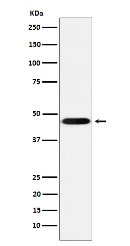
CD16 Rabbit mAb [eJmu]Cat NO.: A93090
Western blot(SDS PAGE) analysis of extracts from THP-1 cell lysate.Using CD16 Rabbit mAb [eJmu]at dilution of 1:1000 incubated at 4℃ over night.
Product information
Protein names :IGFR3; CD16; CD16a; IMD20;
UniProtID :P08637
MASS(da) :29,089
MW(kDa) :42kDa
Form :Liquid
Purification :Affinity-chromatography
Host :Rabbit
Isotype : IgG
sensitivity :Endogenous
Reactivity :Human,Mouse,Rat
- ApplicationDilution
- 免疫印迹(WB)1:1000-2000
- 免疫荧光(ICC/IF)1:100
- The optimal dilutions should be determined by the end user
Specificity :Antibody is produced by immunizing animals with A synthesized peptide derived from human CD16
Storage :Antibody store in 10 mM PBS, 0.5mg/ml BSA, 50% glycerol. Shipped at 4°C. Store at-20°C or -80°C. Products are valid for one natural year of receipt.Avoid repeated freeze / thaw cycles.
WB Positive detected :THP-1 cell lysate.
Function : Receptor for the invariable Fc fragment of immunoglobulin gamma (IgG). Optimally activated upon binding of clustered antigen-IgG complexes displayed on cell surfaces, triggers lysis of antibody-coated cells, a process known as antibody-dependent cellular cytotoxicity (ADCC). Does not bind free monomeric IgG, thus avoiding inappropriate effector cell activation in the absence of antigenic trigger (PubMed:24412922, PubMed:25786175, PubMed:21768335, PubMed:22023369, PubMed:8609432, PubMed:9242542, PubMed:25816339, PubMed:11711607, PubMed:28652325). Mediates IgG effector functions on natural killer (NK) cells. Binds antigen-IgG complexes generated upon infection and triggers NK cell-dependent cytokine production and degranulation to limit viral load and propagation. Involved in the generation of memory-like adaptive NK cells capable to produce high amounts of IFNG and to efficiently eliminate virus-infected cells via ADCC (PubMed:25786175, PubMed:24412922). Regulates NK cell survival and proliferation, in particular by preventing NK cell progenitor apoptosis (PubMed:9916693, PubMed:29967280). Fc-binding subunit that associates with CD247 and/or FCER1G adapters to form functional signaling complexes. Following the engagement of antigen-IgG complexes, triggers phosphorylation of immunoreceptor tyrosine-based activation motif (ITAM)-containing adapters with subsequent activation of phosphatidylinositol 3-kinase signaling and sustained elevation of intracellular calcium that ultimately drive NK cell activation. The ITAM-dependent signaling coupled to receptor phosphorylation by PKC mediates robust intracellular calcium flux that leads to production of pro-inflammatory cytokines, whereas in the absence of receptor phosphorylation it mainly activates phosphatidylinositol 3-kinase signaling leading to cell degranulation (PubMed:2532305, PubMed:1825220, PubMed:23024279). Costimulates NK cells and trigger lysis of target cells independently of IgG binding (PubMed:23006327, PubMed:10318937). Mediates the antitumor activities of therapeutic antibodies. Upon ligation on monocytes triggers TNFA-dependent ADCC of IgG-coated tumor cells (PubMed:27670158). Mediates enhanced ADCC in response to afucosylated IgGs (PubMed:34485821).., (Microbial infection) Involved in Dengue virus pathogenesis via antibody-dependent enhancement (ADE) mechanism. Secondary infection with Dengue virus triggers elevated levels of afucosylated non-neutralizing IgG1s with reactivity to viral envelope/E protein. Viral antigen-IgG1 complexes bind with high affinity to FCGR3A, facilitating virus entry in myeloid cells and subsequent viral replication..
Tissue specificity :Expressed in natural killer cells (at protein level) (PubMed:2526846). Expressed in a subset of circulating monocytes (at protein level) (PubMed:27670158)..
Subcellular locationi :Cell membrane,Single-pass type I membrane protein. Secreted.
IMPORTANT: For western blots, incubate membrane with diluted primary antibody in 1% w/v BSA, 1X TBST at 4°C overnight.


