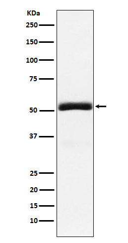
Granulin Rabbit mAb [8D47]Cat NO.: A77779
Western blot(SDS PAGE) analysis of extracts from 293T cell lysate.Using Granulin Rabbit mAb [8D47]at dilution of 1:1000 incubated at 4℃ over night.
Product information
Protein names :Acrogranin; CLN11; GEP; GP88; Granulins; GRN; PCDGF; PEPI; PGRN; Proepithelin; Progranulin;
UniProtID :P28799
MASS(da) :63,544
MW(kDa) :55,75kDa
Form :Liquid
Purification :Affinity-chromatography
Host :Rabbit
Isotype : IgG
sensitivity :Endogenous
Reactivity :Human
- ApplicationDilution
- 免疫印迹(WB)1:1000-2000
- 免疫组化(IHC)1:100
- 免疫荧光(ICC/IF)1:100
- The optimal dilutions should be determined by the end user
Specificity :Antibody is produced by immunizing animals with A synthesized peptide derived from human Granulin
Storage :Antibody store in 10 mM PBS, 0.5mg/ml BSA, 50% glycerol. Shipped at 4°C. Store at-20°C or -80°C. Products are valid for one natural year of receipt.Avoid repeated freeze / thaw cycles.
WB Positive detected :293T cell lysate.
Function : Secreted protein that acts as a key regulator of lysosomal function and as a growth factor involved in inflammation, wound healing and cell proliferation (PubMed:28541286, PubMed:28073925, PubMed:18378771, PubMed:28453791, PubMed:12526812). Regulates protein trafficking to lysosomes and, also the activity of lysosomal enzymes (PubMed:28453791, PubMed:28541286). Facilitates also the acidification of lysosomes, causing degradation of mature CTSD by CTSB (PubMed:28073925). In addition, functions as wound-related growth factor that acts directly on dermal fibroblasts and endothelial cells to promote division, migration and the formation of capillary-like tubule structures (By similarity). Also promotes epithelial cell proliferation by blocking TNF-mediated neutrophil activation preventing release of oxidants and proteases (PubMed:12526812). Moreover, modulates inflammation in neurons by preserving neurons survival, axonal outgrowth and neuronal integrity (PubMed:18378771).., [Granulin-4]: Promotes proliferation of the epithelial cell line A431 in culture., [Granulin-3]: Inhibits epithelial cell proliferation and induces epithelial cells to secrete IL-8.., [Granulin-7]: Stabilizes CTSD through interaction with CTSD leading to maintain its aspartic-type peptidase activity..
Tissue specificity :In myelogenous leukemic cell lines of promonocytic, promyelocytic, and proerythroid lineage, in fibroblasts, and very strongly in epithelial cell lines. Present in inflammatory cells and bone marrow. Highest levels in kidney.
Subcellular locationi :Secreted. Lysosome.
IMPORTANT: For western blots, incubate membrane with diluted primary antibody in 1% w/v BSA, 1X TBST at 4°C overnight.


