
ACVR1 Rabbit mAb [x29Q]Cat NO.: A14153
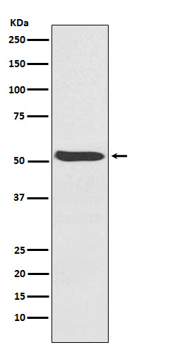
Western blot(SDS PAGE) analysis of extracts from Human fetal heart lysate.Using ACVR1 Rabbit mAb [x29Q]at dilution of 1:1000 incubated at 4℃ over night.
Product information
Protein names :ACTRI; Acvr1; ACVR1A; ACVRLK2; ALK2; FOP; SKR1; TSRI;
UniProtID :Q04771
MASS(da) :57,153
MW(kDa) :57kDa
Form :Liquid
Purification :Affinity-chromatography
Host :Rabbit
Isotype : IgG
sensitivity :Endogenous
Reactivity :Human,Mouse,Rat
- ApplicationDilution
- 免疫印迹(WB)1:1000-2000
- The optimal dilutions should be determined by the end user
Specificity :Antibody is produced by immunizing animals with A synthesized peptide derived from human ACVR1
Storage :Antibody store in 10 mM PBS, 0.5mg/ml BSA, 50% glycerol. Shipped at 4°C. Store at-20°C or -80°C. Products are valid for one natural year of receipt.Avoid repeated freeze / thaw cycles.
WB Positive detected :Human fetal heart lysate.
Function : Bone morphogenetic protein (BMP) type I receptor that is involved in a wide variety of biological processes, including bone, heart, cartilage, nervous, and reproductive system development and regulation (PubMed:20628059, PubMed:22977237). As a type I receptor, forms heterotetrameric receptor complexes with the type II receptors AMHR2, ACVR2A or ACVR2B (PubMed:17911401). Upon binding of ligands such as BMP7 or GDF2/BMP9 to the heteromeric complexes, type II receptors transphosphorylate ACVR1 intracellular domain (PubMed:25354296). In turn, ACVR1 kinase domain is activated and subsequently phosphorylates SMAD1/5/8 proteins that transduce the signal (PubMed:9748228). In addition to its role in mediating BMP pathway-specific signaling, suppresses TGFbeta/activin pathway signaling by interfering with the binding of activin to its type II receptor (PubMed:17911401). Besides canonical SMAD signaling, can activate non-canonical pathways such as p38 mitogen-activated protein kinases/MAPKs (By similarity)..
Tissue specificity :Expressed in normal parenchymal cells, endothelial cells, fibroblasts and tumor-derived epithelial cells..
Subcellular locationi :Membrane,Single-pass type I membrane protein.
IMPORTANT: For western blots, incubate membrane with diluted primary antibody in 1% w/v BSA, 1X TBST at 4°C overnight.
-
About
-
Product
-
Law
-
Literature
-

WeChat scan code follow us