PIM2 Rabbit mAb [eH89]Cat NO.: A21533
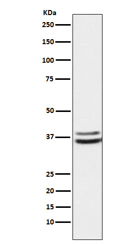
Western blot(SDS PAGE) analysis of extracts from K562 cell lysate.Using PIM2 Rabbit mAb [eH89]at dilution of 1:1000 incubated at 4℃ over night.
Product information
Protein names :PIM2; Pim2h;
UniProtID :Q9P1W9
MASS(da) :34,190
MW(kDa) :34-44kDa
Form :Liquid
Purification :Affinity-chromatography
Host :Rabbit
Isotype : IgG
sensitivity :Endogenous
Reactivity :Human,Mouse,Rat
- ApplicationDilution
- 免疫印迹(WB)1:1000-2000
- The optimal dilutions should be determined by the end user
Specificity :Antibody is produced by immunizing animals with A synthesized peptide derived from PIM2
Storage :Antibody store in 10 mM PBS, 0.5mg/ml BSA, 50% glycerol. Shipped at 4°C. Store at-20°C or -80°C. Products are valid for one natural year of receipt.Avoid repeated freeze / thaw cycles.
WB Positive detected :K562 cell lysate.
Function : Proto-oncogene with serine/threonine kinase activity involved in cell survival and cell proliferation. Exerts its oncogenic activity through: the regulation of MYC transcriptional activity, the regulation of cell cycle progression, the regulation of cap-dependent protein translation and through survival signaling by phosphorylation of a pro-apoptotic protein, BAD. Phosphorylation of MYC leads to an increase of MYC protein stability and thereby an increase transcriptional activity. The stabilization of MYC exerted by PIM2 might explain partly the strong synergism between these 2 oncogenes in tumorigenesis. Regulates cap-dependent protein translation in a mammalian target of rapamycin complex 1 (mTORC1)-independent manner and in parallel to the PI3K-Akt pathway. Mediates survival signaling through phosphorylation of BAD, which induces release of the anti-apoptotic protein Bcl-X(L)/BCL2L1. Promotes cell survival in response to a variety of proliferative signals via positive regulation of the I-kappa-B kinase/NF-kappa-B cascade,this process requires phosphorylation of MAP3K8/COT. Promotes growth factor-independent proliferation by phosphorylation of cell cycle factors such as CDKN1A and CDKN1B. Involved in the positive regulation of chondrocyte survival and autophagy in the epiphyseal growth plate..
Tissue specificity :Highly expressed in hematopoietic tissues, in leukemic and lymphoma cell lines, testis, small intestine, colon and colorectal adenocarcinoma cells. Weakly expressed in normal liver, but highly expressed in hepatocellular carcinoma tissues..
IMPORTANT: For western blots, incubate membrane with diluted primary antibody in 1% w/v BSA, 1X TBST at 4°C overnight.


