
P-MLKL (S345) Rabbit mAb [B5E0]Cat NO.: A42972
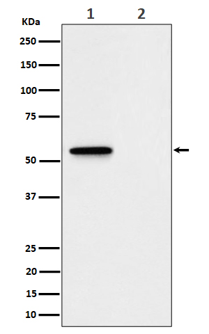
Western blot(SDS PAGE) analysis of extracts from (1) L929 treated with Z-VAD + TNF alpha + SM164 cell lysate; (2) Untreated.Using P-MLKL (S345) Rabbit mAb [B5E0]at dilution of 1:1000 incubated at 4℃ over night.
Product information
Protein names :hMLKL ; Mixed lineage kinase domain like; Mixed lineage kinase domain like protein; Mixed lineage kinase domain like pseudokinase;
UniProtID :Q9D2Y4(Mouse )
MASS(da) :54,317
MW(kDa) :54kDa
Form :Liquid
Purification :Affinity-chromatography
Host :Rabbit
Isotype : IgG
sensitivity :Endogenous
Reactivity :Mouse
- ApplicationDilution
- 免疫印迹(WB)1:1000-2000
- The optimal dilutions should be determined by the end user
Specificity :Antibody is produced by immunizing animals with A synthesized peptide derived from human Phospho-MLKL (S345)
Storage :Antibody store in 10 mM PBS, 0.5mg/ml BSA, 50% glycerol. Shipped at 4°C. Store at-20°C or -80°C. Products are valid for one natural year of receipt.Avoid repeated freeze / thaw cycles.
WB Positive detected :(1) L929 treated with Z-VAD + TNF alpha + SM164 cell lysate; (2) Untreated.
Function : Pseudokinase that plays a key role in TNF-induced necroptosis, a programmed cell death process (PubMed:23835476, PubMed:27321907, PubMed:24012422, PubMed:24019532, PubMed:32200799, PubMed:32296175). Does not have protein kinase activity (PubMed:24012422). Activated following phosphorylation by RIPK3, leading to homotrimerization, localization to the plasma membrane and execution of programmed necrosis characterized by calcium influx and plasma membrane damage (PubMed:23835476, PubMed:27321907, PubMed:24012422, PubMed:24019532). In addition to TNF-induced necroptosis, necroptosis can also take place in the nucleus in response to orthomyxoviruses infection: following ZBP1 activation, which senses double-stranded Z-RNA structures, nuclear RIPK3 catalyzes phosphorylation and activation of MLKL, promoting disruption of the nuclear envelope and leakage of cellular DNA into the cytosol (PubMed:32200799, PubMed:32296175). Binds to highly phosphorylated inositol phosphates such as inositolhexakisphosphate (InsP6) which is essential for its necroptotic function (By similarity)..
Tissue specificity :Highly expressed in thymus, colon, intestine, liver, spleen and lung. Expressed at much lower level in skeletal muscle, heart and kidney. Not detected in brain..
Subcellular locationi :Cytoplasm. Cell membrane. Nucleus.
IMPORTANT: For western blots, incubate membrane with diluted primary antibody in 1% w/v BSA, 1X TBST at 4°C overnight.
-
About
-
Product
-
Law
-
Literature
-

WeChat scan code follow us