
LTK Rabbit mAb [bEz1]Cat NO.: A18365
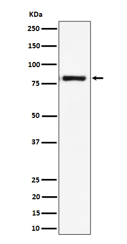
Western blot(SDS PAGE) analysis of extracts from Raji cell lysate.Using LTK Rabbit mAb [bEz1]at dilution of 1:1000 incubated at 4℃ over night.
Product information
Protein names :Ltk; TYK1;
UniProtID :P29376
MASS(da) :91,681
MW(kDa) :80kDa
Form :Liquid
Purification :Affinity-chromatography
Host :Rabbit
Isotype : IgG
sensitivity :Endogenous
Reactivity :Human
- ApplicationDilution
- 免疫印迹(WB)1:1000-2000
- 免疫组化(IHC)1:100
- The optimal dilutions should be determined by the end user
Specificity :Antibody is produced by immunizing animals with A synthesized peptide derived from human LTK
Storage :Antibody store in 10 mM PBS, 0.5mg/ml BSA, 50% glycerol. Shipped at 4°C. Store at-20°C or -80°C. Products are valid for one natural year of receipt.Avoid repeated freeze / thaw cycles.
WB Positive detected :Raji cell lysate.
Function : Receptor with a tyrosine-protein kinase activity (PubMed:10445845, PubMed:20548102, PubMed:30061385). Following activation by ALKAL1 or ALKAL2 ligands at the cell surface, transduces an extracellular signal into an intracellular response (PubMed:30061385, PubMed:34646012). Ligand-binding to the extracellular domain induces tyrosine kinase activation, leading to activation of the mitogen-activated protein kinase (MAPK) pathway (PubMed:20548102). Phosphorylates almost exclusively at the first tyrosine of the Y-x-x-x-Y-Y motif (By similarity). The exact function of this protein is not known,studies with chimeric proteins demonstrate its ability to promote growth and specifically neurite outgrowth, and cell survival (PubMed:9223670, PubMed:18849880). Involved in regulation of the secretory pathway involving endoplasmic reticulum (ER) export sites (ERESs) and ER to Golgi transport (PubMed:20548102)..
Tissue specificity :Expressed in non-hematopoietic cell lines and T- and B-cell lines..
Subcellular locationi :Cell membrane,Single-pass type I membrane protein.
IMPORTANT: For western blots, incubate membrane with diluted primary antibody in 1% w/v BSA, 1X TBST at 4°C overnight.
-
About
-
Product
-
Law
-
Literature
-

WeChat scan code follow us