PLSCR3 Rabbit mAb [H0BI]Cat NO.: A16981
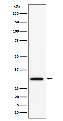
Western blot(SDS PAGE) analysis of extracts from BxPC 3 cell lysate.Using PLSCR3 Rabbit mAb [H0BI]at dilution of 1:1000 incubated at 4℃ over night.
Product information
Protein names :PLS3; Plscr3;
UniProtID :Q9NRY6
MASS(da) :31,648
MW(kDa) :32kDa
Form :Liquid
Purification :Affinity-chromatography
Host :Rabbit
Isotype : IgG
sensitivity :Endogenous
Reactivity :Human,Mouse,Rat
- ApplicationDilution
- 免疫印迹(WB)1:1000-2000
- The optimal dilutions should be determined by the end user
Specificity :Antibody is produced by immunizing animals with A synthesized peptide derived from human PLSCR3
Storage :Antibody store in 10 mM PBS, 0.5mg/ml BSA, 50% glycerol. Shipped at 4°C. Store at-20°C or -80°C. Products are valid for one natural year of receipt.Avoid repeated freeze / thaw cycles.
WB Positive detected :BxPC 3 cell lysate.
Function : Catalyzes calcium-induced ATP-independent rapid bidirectional and non-specific movement of the phospholipids (lipid scrambling or lipid flip-flop) between the inner and outer membrane of the mitochondria (PubMed:14573790, PubMed:17226776, PubMed:18358005, PubMed:29337693, PubMed:31769662). Plays an important role in mitochondrial respiratory function, morphology, and apoptotic response (PubMed:14573790, PubMed:17226776, PubMed:18358005, PubMed:12649167). Mediates the translocation of cardiolipin from the mitochondrial inner membrane to outer membrane enhancing t-Bid induced cytochrome c release and apoptosis (PubMed:14573790, PubMed:17226776, PubMed:18358005). Enhances TNFSF10-induced apoptosis by regulating the distribution of cardiolipin in the mitochondrial membrane resulting in increased release of apoptogenic factors and consequent amplification of the activity of caspases (PubMed:18491232). Regulates cardiolipin de novo biosynthesis and its resynthesis (PubMed:16939411)..
Tissue specificity :Expressed in heart, placenta, lung, liver, skeletal muscle, kidney, pancreas, spleen, thymus, prostate, uterus, small intestine and peripheral blood lymphocytes. Not detected in testis, brain and liver.
Subcellular locationi :Mitochondrion membrane,Single-pass type II membrane protein. Mitochondrion inner membrane,Single-pass type II membrane protein. Nucleus.
IMPORTANT: For western blots, incubate membrane with diluted primary antibody in 1% w/v BSA, 1X TBST at 4°C overnight.


