
AK2 Rabbit mAb [s773]Cat NO.: A28487
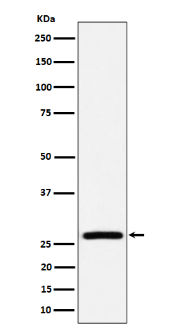
Western blot(SDS PAGE) analysis of extracts from HepG2 cell lysate.Using AK2 Rabbit mAb [s773]at dilution of 1:1000 incubated at 4℃ over night.
Product information
Protein names :Adenylate kinase 2; ADK2; ak2;
UniProtID :P54819
MASS(da) :26,478
MW(kDa) :26kDa
Form :Liquid
Purification :Affinity-chromatography
Host :Rabbit
Isotype : IgG
sensitivity :Endogenous
Reactivity :Human,Mouse,Rat
- ApplicationDilution
- 免疫印迹(WB)1:1000-2000
- 免疫组化(IHC)1:100
- 免疫荧光(ICC/IF)1:100
- The optimal dilutions should be determined by the end user
Specificity :Antibody is produced by immunizing animals with A synthesized peptide derived from human AK2
Storage :Antibody store in 10 mM PBS, 0.5mg/ml BSA, 50% glycerol. Shipped at 4°C. Store at-20°C or -80°C. Products are valid for one natural year of receipt.Avoid repeated freeze / thaw cycles.
WB Positive detected :HepG2 cell lysate.
Function : Catalyzes the reversible transfer of the terminal phosphate group between ATP and AMP. Plays an important role in cellular energy homeostasis and in adenine nucleotide metabolism. Adenylate kinase activity is critical for regulation of the phosphate utilization and the AMP de novo biosynthesis pathways. Plays a key role in hematopoiesis..
Tissue specificity :Present in most tissues. Present at high level in heart, liver and kidney, and at low level in brain, skeletal muscle and skin. Present in thrombocytes but not in erythrocytes, which lack mitochondria. Present in all nucleated cell populations from blood, while AK1 is mostly absent. In spleen and lymph nodes, mononuclear cells lack AK1, whereas AK2 is readily detectable. These results indicate that leukocytes may be susceptible to defects caused by the lack of AK2, as they do not express AK1 in sufficient amounts to compensate for the AK2 functional deficits (at protein level)..
Subcellular locationi :Mitochondrion intermembrane space.
IMPORTANT: For western blots, incubate membrane with diluted primary antibody in 1% w/v BSA, 1X TBST at 4°C overnight.
-
About
-
Product
-
Law
-
Literature
-

WeChat scan code follow us