TAOK1 Rabbit mAb [owl2]Cat NO.: A82066
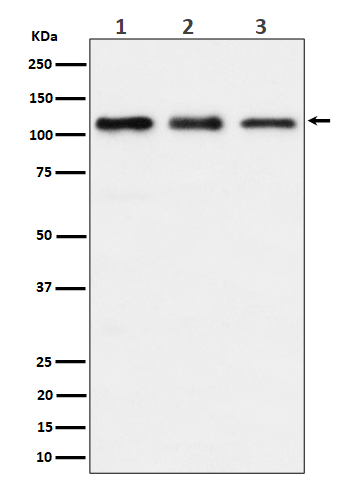
Western blot(SDS PAGE) analysis of extracts from (1) HeLa cell lysate; (2) RAW 264.7 cell lysate; (3) C6 cell lysate.Using TAOK1 Rabbit mAb [owl2]at dilution of 1:1000 incubated at 4℃ over night.
Product information
Protein names :hKFC B; MAP3K16; MARK kinase; MARKK; PSK2; STE20 like kinase; Taok1;
UniProtID :Q7L7X3
MASS(da) :116,070
MW(kDa) :116kDa
Form :Liquid
Purification :Affinity-chromatography
Host :Rabbit
Isotype : IgG
sensitivity :Endogenous
Reactivity :Human,Mouse,Rat
- ApplicationDilution
- 免疫印迹(WB)1:1000-2000
- The optimal dilutions should be determined by the end user
Specificity :Antibody is produced by immunizing animals with A synthesized peptide derived from human TAOK1
Storage :Antibody store in 10 mM PBS, 0.5mg/ml BSA, 50% glycerol. Shipped at 4°C. Store at-20°C or -80°C. Products are valid for one natural year of receipt.Avoid repeated freeze / thaw cycles.
WB Positive detected :(1) HeLa cell lysate; (2) RAW 264.7 cell lysate; (3) C6 cell lysate.
Function : Serine/threonine-protein kinase involved in various processes such as p38/MAPK14 stress-activated MAPK cascade, DNA damage response and regulation of cytoskeleton stability. Phosphorylates MAP2K3, MAP2K6 and MARK2. Acts as an activator of the p38/MAPK14 stress-activated MAPK cascade by mediating phosphorylation and subsequent activation of the upstream MAP2K3 and MAP2K6 kinases. Involved in G-protein coupled receptor signaling to p38/MAPK14. In response to DNA damage, involved in the G2/M transition DNA damage checkpoint by activating the p38/MAPK14 stress-activated MAPK cascade, probably by mediating phosphorylation of MAP2K3 and MAP2K6. Acts as a regulator of cytoskeleton stability by phosphorylating 'Thr-208' of MARK2, leading to activate MARK2 kinase activity and subsequent phosphorylation and detachment of MAPT/TAU from microtubules. Also acts as a regulator of apoptosis: regulates apoptotic morphological changes, including cell contraction, membrane blebbing and apoptotic bodies formation via activation of the MAPK8/JNK cascade..
Tissue specificity :Highly expressed in the testis, and to a lower extent also expressed in brain, placenta, colon and skeletal muscle..
Subcellular locationi :Cytoplasm.
IMPORTANT: For western blots, incubate membrane with diluted primary antibody in 1% w/v BSA, 1X TBST at 4°C overnight.


