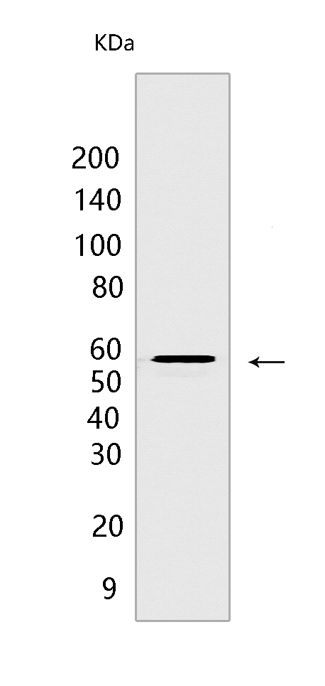
Calcineurin A Rabbit mAb [K4EU]Cat NO.: A85849
Western blot(SDS PAGE) analysis of extracts from Wild-type HAP1 cells.Using Calcineurin ARabbit mAb [K4EU] at dilution of 1:1000 incubated at 4℃ over night.
Product information
Protein names :PPP3CA,CALNA,CNA,PP2BA_HUMAN,Protein phosphatase 3 catalytic subunit alpha
UniProtID :Q08209
MASS(da) :58,688
MW(kDa) :59/32 kDa
Form :Liquid
Purification :Protein A purification
Host :Rabbit
Isotype :IgG
sensitivity :Endogenous
Reactivity :Human,Mouse,Rat
- ApplicationDilution
- 免疫印迹(WB)1:1000-2000
- 免疫组化(IHC)1:100
- The optimal dilutions should be determined by the end user
Specificity :Antibody is produced by immunizing animals with a synthetic peptide at the sequence of human Calcineurin A
Storage :Antibody store in 10 mM PBS, 0.5mg/ml BSA, 50% glycerol. Shipped at 4°C. Store at-20°C or -80°C. Products are valid for one natural year of receipt.Avoid repeated freeze / thaw cycles.
WB Positive detected :Wild-type HAP1 cells
Function : Calcium-dependent, calmodulin-stimulated protein phosphatase which plays an essential role in the transduction of intracellular Ca(2+)-mediated signals (PubMed:15671020, PubMed:18838687, PubMed:19154138, PubMed:23468591, PubMed:30254215). Many of the substrates contain a PxIxIT motif and/or a LxVP motif (PubMed:17498738, PubMed:17502104, PubMed:22343722, PubMed:23468591, PubMed:27974827). In response to increased Ca(2+) levels, dephosphorylates and activates phosphatase SSH1 which results in cofilin dephosphorylation (PubMed:15671020). In response to increased Ca(2+) levels following mitochondrial depolarization, dephosphorylates DNM1L inducing DNM1L translocation to the mitochondrion (PubMed:18838687). Positively regulates the CACNA1B/CAV2.2-mediated Ca(2+) release probability at hippocampal neuronal soma and synaptic terminals (By similarity). Dephosphorylates heat shock protein HSPB1 (By similarity). Dephosphorylates and activates transcription factor NFATC1 (PubMed:19154138). In response to increased Ca(2+) levels, regulates NFAT-mediated transcription probably by dephosphorylating NFAT and promoting its nuclear translocation (PubMed:26248042). Dephosphorylates and inactivates transcription factor ELK1 (PubMed:19154138). Dephosphorylates DARPP32 (PubMed:19154138). May dephosphorylate CRTC2 at 'Ser-171' resulting in CRTC2 dissociation from 14-3-3 proteins (PubMed:30611118). Dephosphorylates transcription factor TFEB at 'Ser-211' following Coxsackievirus B3 infection, promoting nuclear translocation (PubMed:33691586). Required for postnatal development of the nephrogenic zone and superficial glomeruli in the kidneys, cell cycle homeostasis in the nephrogenic zone, and ultimately normal kidney function (By similarity). Plays a role in intracellular AQP2 processing and localization to the apical membrane in the kidney, may thereby be required for efficient kidney filtration (By similarity). Required for secretion of salivary enzymes amylase, peroxidase, lysozyme and sialic acid via formation of secretory vesicles in the submandibular glands (By similarity). Required for calcineurin activity and homosynaptic depotentiation in the hippocampus (By similarity). Required for normal differentiation and survival of keratinocytes and therefore required for epidermis superstructure formation (By similarity). Positively regulates osteoblastic bone formation, via promotion of osteoblast differentiation (By similarity). Positively regulates osteoclast differentiation, potentially via NFATC1 signaling (By similarity). May play a role in skeletal muscle fiber type specification, potentially via NFATC1 signaling (By similarity). Negatively regulates MAP3K14/NIK signaling via inhibition of nuclear translocation of the transcription factors RELA and RELB (By similarity). Required for antigen-specific T-cell proliferation response (By similarity)..
Tissue specificity :Expressed in keratinocytes (at protein level) (PubMed:29043977). Expressed in lymphoblasts (at protein level) (PubMed:30254215)..
Subcellular locationi :Cytoplasm. Cell membrane,Peripheral membrane protein. Cell membrane, sarcolemma. Cytoplasm, myofibril, sarcomere, Z line. Cell projection, dendritic spine.
IMPORTANT: For western blots, incubate membrane with diluted primary antibody in 1% w/v BSA, 1X TBST at 4°C overnight.


