HDAC9 Rabbit mAb [7L4X]Cat NO.: A65084
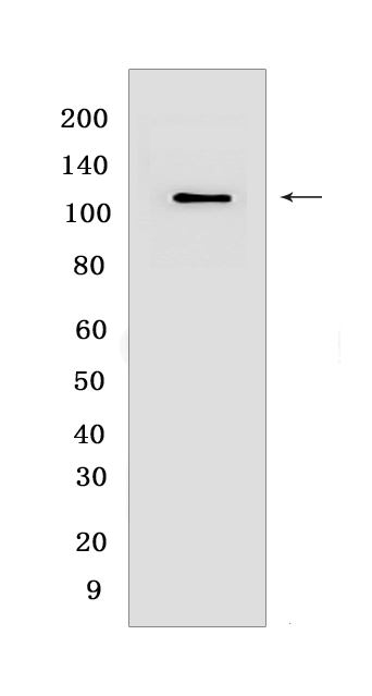
Western blot(SDS PAGE) analysis of extracts from HepG2 cells.Using HDAC9Rabbit mAb [7L4X] at dilution of 1:1000 incubated at 4℃ over night.
Product information
Protein names :HDAC9,HDAC7,HDAC7B,HDRP,KIAA0744,MITR,HDAC9_HUMAN,Histone deacetylase 9
UniProtID :Q9UKV0
MASS(da) :111,297
MW(kDa) :110 kDa
Form :Liquid
Purification :Protein A purification
Host :Rabbit
Isotype :IgG
sensitivity :Endogenous
Reactivity :Human,Mouse,Rat
- ApplicationDilution
- 免疫印迹(WB)1:1000-2000
- 免疫组化(IHC)1:100
- 免疫荧光(ICC/IF) 1:100
- The optimal dilutions should be determined by the end user
Specificity :Antibody is produced by immunizing animals with a synthetic peptide at the sequence of human HDAC9
Storage :Antibody store in 10 mM PBS, 0.5mg/ml BSA, 50% glycerol. Shipped at 4°C. Store at-20°C or -80°C. Products are valid for one natural year of receipt.Avoid repeated freeze / thaw cycles.
WB Positive detected :HepG2 cells
Function : Responsible for the deacetylation of lysine residues on the N-terminal part of the core histones (H2A, H2B, H3 and H4). Histone deacetylation gives a tag for epigenetic repression and plays an important role in transcriptional regulation, cell cycle progression and developmental events. Represses MEF2-dependent transcription.., Isoform 3 lacks active site residues and therefore is catalytically inactive. Represses MEF2-dependent transcription by recruiting HDAC1 and/or HDAC3. Seems to inhibit skeletal myogenesis and to be involved in heart development. Protects neurons from apoptosis, both by inhibiting JUN phosphorylation by MAPK10 and by repressing JUN transcription via HDAC1 recruitment to JUN promoter.
Tissue specificity :Broadly expressed, with highest levels in brain, heart, muscle and testis. Isoform 3 is present in human bladder carcinoma cells (at protein level)..
Subcellular locationi :Nucleus.
IMPORTANT: For western blots, incubate membrane with diluted primary antibody in 1% w/v BSA, 1X TBST at 4°C overnight.


