
MERTK Rabbit mAb [55GH]Cat NO.: A13686
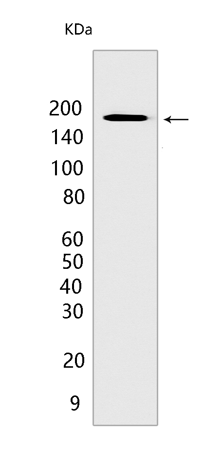
Western blot(SDS PAGE) analysis of extracts from HEK-293 cells.Using MERTKRabbit mAb [55GH] at dilution of 1:1000 incubated at 4℃ over night.
Product information
Protein names :MERTK,MER,MERTK_HUMAN,Tyrosine-protein kinase Mer
UniProtID :Q12866
MASS(da) :110,249
MW(kDa) :180 kDa
Form :Liquid
Purification :Protein A purification
Host :Rabbit
Isotype :IgG
sensitivity :Endogenous
Reactivity :Human
- ApplicationDilution
- 免疫印迹(WB)1:1000-2000
- 免疫组化(IHC)1:100
- The optimal dilutions should be determined by the end user
Specificity :Antibody is produced by immunizing animals with a synthetic peptide at the sequence of human MERTK
Storage :Antibody store in 10 mM PBS, 0.5mg/ml BSA, 50% glycerol. Shipped at 4°C. Store at-20°C or -80°C. Products are valid for one natural year of receipt.Avoid repeated freeze / thaw cycles.
WB Positive detected : HEK-293 cells
Function : Receptor tyrosine kinase that transduces signals from the extracellular matrix into the cytoplasm by binding to several ligands including LGALS3, TUB, TULP1 or GAS6. Regulates many physiological processes including cell survival, migration, differentiation, and phagocytosis of apoptotic cells (efferocytosis). Ligand binding at the cell surface induces autophosphorylation of MERTK on its intracellular domain that provides docking sites for downstream signaling molecules. Following activation by ligand, interacts with GRB2 or PLCG2 and induces phosphorylation of MAPK1, MAPK2, FAK/PTK2 or RAC1. MERTK signaling plays a role in various processes such as macrophage clearance of apoptotic cells, platelet aggregation, cytoskeleton reorganization and engulfment (PubMed:32640697). Functions in the retinal pigment epithelium (RPE) as a regulator of rod outer segments fragments phagocytosis. Plays also an important role in inhibition of Toll-like receptors (TLRs)-mediated innate immune response by activating STAT1, which selectively induces production of suppressors of cytokine signaling SOCS1 and SOCS3..
Tissue specificity :Not expressed in normal B- and T-lymphocytes but is expressed in numerous neoplastic B- and T-cell lines. Highly expressed in testis, ovary, prostate, lung, and kidney, with lower expression in spleen, small intestine, colon, and liver.
Subcellular locationi :Cell membrane,Single-pass type I membrane protein.
IMPORTANT: For western blots, incubate membrane with diluted primary antibody in 1% w/v BSA, 1X TBST at 4°C overnight.
-
About
-
Product
-
Law
-
Literature
-

WeChat scan code follow us