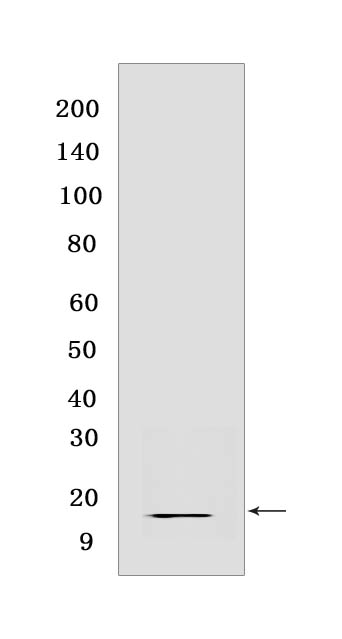
Midkine Rabbit mAb [DPS4]Cat NO.: A81301
Western blot(SDS PAGE) analysis of extracts from Human liver carcinoma.Using MidkineRabbit mAb [DPS4] at dilution of 1:1000 incubated at 4℃ over night.
Product information
Protein names :MDK,MK1,NEGF2,MK_HUMAN,Midkine
UniProtID :P21741
MASS(da) :15,585
MW(kDa) :16 kDa
Form :Liquid
Purification :Protein A purification
Host :Rabbit
Isotype :IgG
sensitivity :Endogenous
Reactivity :Human
- ApplicationDilution
- 免疫印迹(WB)1:1000-2000
- 免疫组化(IHC)1:100
- 免疫荧光(ICC/IF) 1:100
- The optimal dilutions should be determined by the end user
Specificity :Antibody is produced by immunizing animals with a synthetic peptide at the sequence of human Midkine
Storage :Antibody store in 10 mM PBS, 0.5mg/ml BSA, 50% glycerol. Shipped at 4°C. Store at-20°C or -80°C. Products are valid for one natural year of receipt.Avoid repeated freeze / thaw cycles.
WB Positive detected :Human liver carcinoma
Function : Secreted protein that functions as cytokine and growth factor and mediates its signal through cell-surface proteoglycan and non-proteoglycan receptors (PubMed:18469519, PubMed:12573468, PubMed:12122009, PubMed:10212223, PubMed:24458438, PubMed:15466886, PubMed:12084985, PubMed:10772929). Binds cell-surface proteoglycan receptors via their chondroitin sulfate (CS) groups (PubMed:12084985, PubMed:10212223). Thereby regulates many processes like inflammatory response, cell proliferation, cell adhesion, cell growth, cell survival, tissue regeneration, cell differentiation and cell migration (PubMed:12573468, PubMed:12122009, PubMed:10212223, PubMed:10683378, PubMed:24458438, PubMed:22323540, PubMed:12084985, PubMed:15466886, PubMed:10772929). Participates in inflammatory processes by exerting two different activities. Firstly, mediates neutrophils and macrophages recruitment to the sites of inflammation both by direct action by cooperating namely with ITGB2 via LRP1 and by inducing chemokine expression (PubMed:10683378, PubMed:24458438). This inflammation can be accompanied by epithelial cell survival and smooth muscle cell migration after renal and vessel damage, respectively (PubMed:10683378). Secondly, suppresses the development of tolerogenic dendric cells thereby inhibiting the differentiation of regulatory T cells and also promote T cell expansion through NFAT signaling and Th1 cell differentiation (PubMed:22323540). Promotes tissue regeneration after injury or trauma. After heart damage negatively regulates the recruitment of inflammatory cells and mediates cell survival through activation of anti-apoptotic signaling pathways via MAPKs and AKT pathways through the activation of angiogenesis (By similarity). Also facilitates liver regeneration as well as bone repair by recruiting macrophage at trauma site and by promoting cartilage development by facilitating chondrocyte differentiation (By similarity). Plays a role in brain by promoting neural precursor cells survival and growth through interaction with heparan sulfate proteoglycans (By similarity). Binds PTPRZ1 and promotes neuronal migration and embryonic neurons survival (PubMed:10212223). Binds SDC3 or GPC2 and mediates neurite outgrowth and cell adhesion (PubMed:12084985, PubMed:1768439). Binds chondroitin sulfate E and heparin leading to inhibition of neuronal cell adhesion induced by binding with GPC2 (PubMed:12084985). Binds CSPG5 and promotes elongation of oligodendroglial precursor-like cells (By similarity). Also binds ITGA6:ITGB1 complex,this interaction mediates MDK-induced neurite outgrowth (PubMed:15466886, PubMed:1768439). Binds LRP1,promotes neuronal survival (PubMed:10772929). Binds ITGA4:ITGB1 complex,this interaction mediates MDK-induced osteoblast cells migration through PXN phosphorylation (PubMed:15466886). Binds anaplastic lymphoma kinase (ALK) which induces ALK activation and subsequent phosphorylation of the insulin receptor substrate (IRS1), followed by the activation of mitogen-activated protein kinase (MAPK) and PI3-kinase, and the induction of cell proliferation (PubMed:12122009). Promotes epithelial to mesenchymal transition through interaction with NOTCH2 (PubMed:18469519). During arteriogenesis, plays a role in vascular endothelial cell proliferation by inducing VEGFA expression and release which in turn induces nitric oxide synthase expression. Moreover activates vasodilation through nitric oxide synthase activation (By similarity). Negatively regulates bone formation in response to mechanical load by inhibiting Wnt/beta-catenin signaling in osteoblasts (By similarity). In addition plays a role in hippocampal development, working memory, auditory response, early fetal adrenal gland development and the female reproductive system (By similarity)..
Tissue specificity :Expressed in various tumor cell lines. In insulinoma tissue predominantly expressed in precancerous lesions..
Subcellular locationi :Secreted.
IMPORTANT: For western blots, incubate membrane with diluted primary antibody in 1% w/v BSA, 1X TBST at 4°C overnight.


