
PODXL Rabbit mAb [4K0F]Cat NO.: A14265
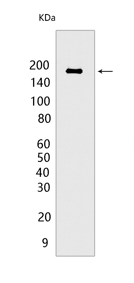
Western blot(SDS PAGE) analysis of extracts from HCT 116 CELLS .Using PODXLRabbit mAb [4K0F] at dilution of 1:1000 incubated at 4℃ over night.
Product information
Protein names :PODXL,PCLP,PCLP1,PODXL_HUMAN,Podocalyxin
UniProtID :O00592
MASS(da) :58,635
MW(kDa) :165 kDa
Form :Liquid
Purification :Protein A purification
Host :Rabbit
Isotype :IgG
sensitivity :Endogenous
Reactivity :Human
- ApplicationDilution
- 免疫印迹(WB)1:1000-2000
- 免疫组化(IHC)1:100
- 免疫荧光(ICC/IF) 1:100
- The optimal dilutions should be determined by the end user
Specificity :Antibody is produced by immunizing animals with a synthetic peptide at the sequence of human PODXL
Storage :Antibody store in 10 mM PBS, 0.5mg/ml BSA, 50% glycerol. Shipped at 4°C. Store at-20°C or -80°C. Products are valid for one natural year of receipt.Avoid repeated freeze / thaw cycles.
WB Positive detected :HCT 116 CELLS
Function : Involved in the regulation of both adhesion and cell morphology and cancer progression. Functions as an anti-adhesive molecule that maintains an open filtration pathway between neighboring foot processes in the podocyte by charge repulsion. Acts as a pro-adhesive molecule, enhancing the adherence of cells to immobilized ligands, increasing the rate of migration and cell-cell contacts in an integrin-dependent manner. Induces the formation of apical actin-dependent microvilli. Involved in the formation of a preapical plasma membrane subdomain to set up initial epithelial polarization and the apical lumen formation during renal tubulogenesis. Plays a role in cancer development and aggressiveness by inducing cell migration and invasion through its interaction with the actin-binding protein EZR. Affects EZR-dependent signaling events, leading to increased activities of the MAPK and PI3K pathways in cancer cells..
Tissue specificity :Glomerular epithelium cell (podocyte).
Subcellular locationi :Apical cell membrane. Cell projection, lamellipodium. Cell projection, filopodium. Cell projection, ruffle. Cell projection, microvillus. Membrane raft. Membrane,Single-pass type I membrane protein.
IMPORTANT: For western blots, incubate membrane with diluted primary antibody in 1% w/v BSA, 1X TBST at 4°C overnight.
-
About
-
Product
-
Law
-
Literature
-

WeChat scan code follow us