
PKD2 Rabbit mAb [SM5M]Cat NO.: A17778
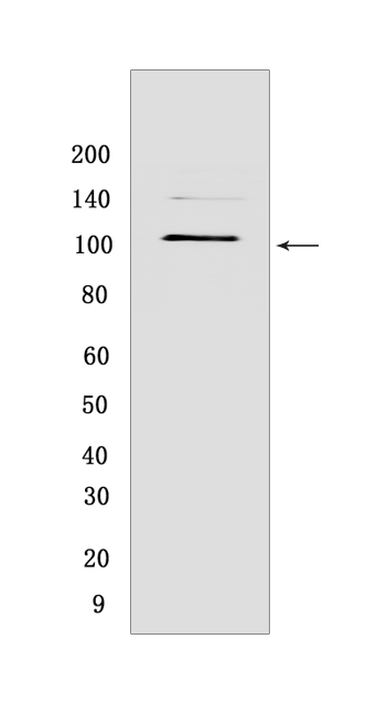
Western blot(SDS PAGE) analysis of extracts from HeLa cells .Using PKD2Rabbit mAb [SM5M] at dilution of 1:1000 incubated at 4℃ over night.
Product information
Protein names :PKD2,TRPP2,PKD2_HUMAN,Polycystin-2
UniProtID :Q13563
MASS(da) :109,691
MW(kDa) :105 kDa
Form :Liquid
Purification :Protein A purification
Host :Rabbit
Isotype :IgG
sensitivity :Endogenous
Reactivity :Human,Mouse
- ApplicationDilution
- 免疫印迹(WB)1:1000-2000
- 免疫组化(IHC)1:100
- 免疫荧光(ICC/IF) 1:100
- The optimal dilutions should be determined by the end user
Specificity :Antibody is produced by immunizing animals with a synthetic peptide at the sequence of human PKD2
Storage :Antibody store in 10 mM PBS, 0.5mg/ml BSA, 50% glycerol. Shipped at 4°C. Store at-20°C or -80°C. Products are valid for one natural year of receipt.Avoid repeated freeze / thaw cycles.
WB Positive detected :HeLa cells
Function : Component of a heteromeric calcium-permeable ion channel formed by PKD1 and PKD2 that is activated by interaction between PKD1 and a Wnt family member, such as WNT3A and WNT9B (PubMed:27214281). Can also form a functional, homotetrameric ion channel (PubMed:29899465). Functions as a cation channel involved in fluid-flow mechanosensation by the primary cilium in renal epithelium (PubMed:18695040). Functions as outward-rectifying K(+) channel, but is also permeable to Ca(2+), and to a much lesser degree also to Na(+) (PubMed:11854751, PubMed:15692563, PubMed:27071085, PubMed:27991905). May contribute to the release of Ca(2+) stores from the endoplasmic reticulum (PubMed:11854751, PubMed:20881056). Together with TRPV4, forms mechano- and thermosensitive channels in cilium (PubMed:18695040). PKD1 and PKD2 may function through a common signaling pathway that is necessary to maintain the normal, differentiated state of renal tubule cells. Acts as a regulator of cilium length, together with PKD1. The dynamic control of cilium length is essential in the regulation of mechanotransductive signaling. The cilium length response creates a negative feedback loop whereby fluid shear-mediated deflection of the primary cilium, which decreases intracellular cAMP, leads to cilium shortening and thus decreases flow-induced signaling. Also involved in left-right axis specification via its role in sensing nodal flow,forms a complex with PKD1L1 in cilia to facilitate flow detection in left-right patterning. Detection of asymmetric nodal flow gives rise to a Ca(2+) signal that is required for normal, asymmetric expression of genes involved in the specification of body left-right laterality (By similarity)..
Tissue specificity :Detected in fetal and adult kidney (PubMed:10770959). Detected at the thick ascending limb of the loop of Henle, at distal tubules, including the distal convoluted tubule and cortical collecting tubules, with weak staining of the collecting duct (PubMed:10770959). Detected on placenta syncytiotrophoblasts (at protein level) (PubMed:26269590). Strongly expressed in ovary, fetal and adult kidney, testis, and small intestine. Not detected in peripheral leukocytes..
Subcellular locationi :Cell projection, cilium membrane,Multi-pass membrane protein. Endoplasmic reticulum membrane,Multi-pass membrane protein. Cell membrane,Multi-pass membrane protein. Basolateral cell membrane. Cytoplasmic vesicle membrane. Golgi apparatus.
IMPORTANT: For western blots, incubate membrane with diluted primary antibody in 1% w/v BSA, 1X TBST at 4°C overnight.
-
About
-
Product
-
Law
-
Literature
-

WeChat scan code follow us