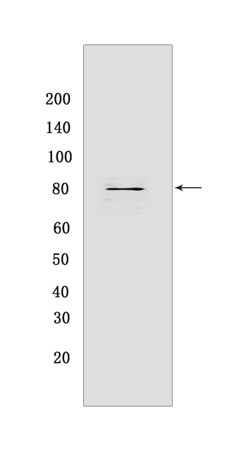
NAK/TBK1 Rabbit mAb [SS03]Cat NO.: A56414
Western blot(SDS PAGE) analysis of extracts from HepG2 cells.Using NAK/TBK1Rabbit mAb [SS03] at dilution of 1:1000 incubated at 4℃ over night.
Product information
Protein names :TBK1,NAK,TBK1_HUMAN,Serine/threonine-protein kinase TBK1
UniProtID :Q9UHD2
MASS(da) :83,642
MW(kDa) :84 kDa
Form :Liquid
Purification :Protein A purification
Host :Rabbit
Isotype :IgG
sensitivity :Endogenous
Reactivity :Human,Mouse
- ApplicationDilution
- 免疫印迹(WB)1:1000-2000
- 免疫组化(IHC)1:100
- The optimal dilutions should be determined by the end user
Specificity :Antibody is produced by immunizing animals with a synthetic peptide at the sequence of human NAK/TBK1
Storage :Antibody store in 10 mM PBS, 0.5mg/ml BSA, 50% glycerol. Shipped at 4°C. Store at-20°C or -80°C. Products are valid for one natural year of receipt.Avoid repeated freeze / thaw cycles.
WB Positive detected :HepG2 cells
Function : Serine/threonine kinase that plays an essential role in regulating inflammatory responses to foreign agents (PubMed:12692549, PubMed:14703513, PubMed:18583960, PubMed:12702806, PubMed:15367631, PubMed:10581243, PubMed:11839743, PubMed:15485837, PubMed:21138416, PubMed:25636800, PubMed:23453971, PubMed:23453972, PubMed:23746807, PubMed:26611359, PubMed:32404352). Following activation of toll-like receptors by viral or bacterial components, associates with TRAF3 and TANK and phosphorylates interferon regulatory factors (IRFs) IRF3 and IRF7 as well as DDX3X (PubMed:12692549, PubMed:14703513, PubMed:18583960, PubMed:12702806, PubMed:15367631, PubMed:25636800). This activity allows subsequent homodimerization and nuclear translocation of the IRFs leading to transcriptional activation of pro-inflammatory and antiviral genes including IFNA and IFNB (PubMed:12702806, PubMed:15367631, PubMed:25636800, PubMed:32972995). In order to establish such an antiviral state, TBK1 form several different complexes whose composition depends on the type of cell and cellular stimuli (PubMed:23453971, PubMed:23453972, PubMed:23746807). Plays a key role in IRF3 activation: acts by first phosphorylating innate adapter proteins MAVS, STING1 and TICAM1 on their pLxIS motif, leading to recruitment of IRF3, thereby licensing IRF3 for phosphorylation by TBK1 (PubMed:25636800, PubMed:30842653). Phosphorylated IRF3 dissociates from the adapter proteins, dimerizes, and then enters the nucleus to induce expression of interferons (PubMed:25636800). Thus, several scaffolding molecules including FADD, TRADD, MAVS, AZI2, TANK or TBKBP1/SINTBAD can be recruited to the TBK1-containing-complexes (PubMed:21931631). Under particular conditions, functions as a NF-kappa-B effector by phosphorylating NF-kappa-B inhibitor alpha/NFKBIA, IKBKB or RELA to translocate NF-Kappa-B to the nucleus (PubMed:10783893, PubMed:15489227). Restricts bacterial proliferation by phosphorylating the autophagy receptor OPTN/Optineurin on 'Ser-177', thus enhancing LC3 binding affinity and antibacterial autophagy (PubMed:21617041). Phosphorylates SMCR8 component of the C9orf72-SMCR8 complex, promoting autophagosome maturation (PubMed:27103069). Phosphorylates ATG8 proteins MAP1LC3C and GABARAPL2, thereby preventing their delipidation and premature removal from nascent autophagosomes (PubMed:31709703). Phosphorylates and activates AKT1 (PubMed:21464307). Seems to play a role in energy balance regulation by sustaining a state of chronic, low-grade inflammation in obesity, wich leads to a negative impact on insulin sensitivity (By similarity). Attenuates retroviral budding by phosphorylating the endosomal sorting complex required for transport-I (ESCRT-I) subunit VPS37C (PubMed:21270402). Phosphorylates Borna disease virus (BDV) P protein (PubMed:16155125). Plays an essential role in the TLR3- and IFN-dependent control of herpes virus HSV-1 and HSV-2 infections in the central nervous system (PubMed:22851595)..
Tissue specificity :Ubiquitous with higher expression in testis. Expressed in the ganglion cells, nerve fiber layer and microvasculature of the retina..
Subcellular locationi :Cytoplasm.
IMPORTANT: For western blots, incubate membrane with diluted primary antibody in 1% w/v BSA, 1X TBST at 4°C overnight.


