
VAV3 Rabbit mAb [FZ3D]Cat NO.: A44947
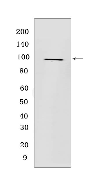
Western blot(SDS PAGE) analysis of extracts from Jurkat cells .Using VAV3Rabbit mAb [FZ3D] at dilution of 1:1000 incubated at 4℃ over night.
Product information
Protein names :VAV3,VAV3_HUMAN,Guanine nucleotide exchange factor VAV3
UniProtID :Q9UKW4
MASS(da) :97,776
MW(kDa) :98 kDa
Form :Liquid
Purification :Protein A purification
Host :Rabbit
Isotype :IgG
sensitivity :Endogenous
Reactivity :Human
- ApplicationDilution
- 免疫印迹(WB)1:1000-2000
- The optimal dilutions should be determined by the end user
Specificity :Antibody is produced by immunizing animals with a synthetic peptide at the sequence of human VAV3
Storage :Antibody store in 10 mM PBS, 0.5mg/ml BSA, 50% glycerol. Shipped at 4°C. Store at-20°C or -80°C. Products are valid for one natural year of receipt.Avoid repeated freeze / thaw cycles.
WB Positive detected :Jurkat cells
Function : Exchange factor for GTP-binding proteins RhoA, RhoG and, to a lesser extent, Rac1. Binds physically to the nucleotide-free states of those GTPases. Plays an important role in angiogenesis. Its recruitment by phosphorylated EPHA2 is critical for EFNA1-induced RAC1 GTPase activation and vascular endothelial cell migration and assembly (By similarity). May be important for integrin-mediated signaling, at least in some cell types. In osteoclasts, along with SYK tyrosine kinase, required for signaling through integrin alpha-v/beta-1 (ITAGV-ITGB1), a crucial event for osteoclast proper cytoskeleton organization and function. This signaling pathway involves RAC1, but not RHO, activation. Necessary for proper wound healing. In the course of wound healing, required for the phagocytotic cup formation preceding macrophage phagocytosis of apoptotic neutrophils. Responsible for integrin beta-2 (ITGB2)-mediated macrophage adhesion and, to a lesser extent, contributes to beta-3 (ITGB3)-mediated adhesion. Does not affect integrin beta-1 (ITGB1)-mediated adhesion (By similarity)..
Tissue specificity :Isoform 1 and isoform 3 are widely expressed,both are expressed at very low levels in skeletal muscle. In keratinocytes, isoform 1 is less abundant than isoform 3. Isoform 3 is detected at very low levels, if any, in adrenal gland, bone marrow, spleen, fetal brain and spinal chord,in these tissues, isoform 1 is readily detectable..
IMPORTANT: For western blots, incubate membrane with diluted primary antibody in 1% w/v BSA, 1X TBST at 4°C overnight.
-
About
-
Product
-
Law
-
Literature
-

WeChat scan code follow us