
Eph receptor B4/HTK Rabbit mAb [SFHD]Cat NO.: A46874
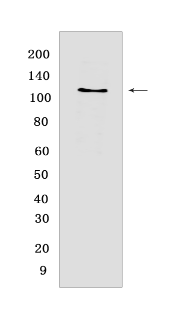
Western blot(SDS PAGE) analysis of extracts from HuvEc cells.Using Eph receptor B4/HTKRabbit mAb [SFHD] at dilution of 1:1000 incubated at 4℃ over night.
Product information
Protein names :EPHB4,HTK,MYK1,TYRO11,EPHB4_HUMAN,Ephrin type-B receptor 4
UniProtID :P54760
MASS(da) :108,270
MW(kDa) :120 kDa
Form :Liquid
Purification :Protein A purification
Host :Rabbit
Isotype :IgG
sensitivity :Endogenous
Reactivity :Human,Mouse
- ApplicationDilution
- 免疫印迹(WB)1:1000-2000
- 免疫组化(IHC)1:100
- The optimal dilutions should be determined by the end user
Specificity :Antibody is produced by immunizing animals with a synthetic peptide at the sequence of human Eph receptor B4/HTK
Storage :Antibody store in 10 mM PBS, 0.5mg/ml BSA, 50% glycerol. Shipped at 4°C. Store at-20°C or -80°C. Products are valid for one natural year of receipt.Avoid repeated freeze / thaw cycles.
WB Positive detected :HuvEc cells
Function : Receptor tyrosine kinase which binds promiscuously transmembrane ephrin-B family ligands residing on adjacent cells, leading to contact-dependent bidirectional signaling into neighboring cells. The signaling pathway downstream of the receptor is referred to as forward signaling while the signaling pathway downstream of the ephrin ligand is referred to as reverse signaling. Together with its cognate ligand/functional ligand EFNB2 it is involved in the regulation of cell adhesion and migration, and plays a central role in heart morphogenesis, angiogenesis and blood vessel remodeling and permeability. EPHB4-mediated forward signaling controls cellular repulsion and segregation from EFNB2-expressing cells..
Tissue specificity :Abundantly expressed in placenta but also detected in kidney, liver, lung, pancreas, skeletal muscle and heart. Expressed in primitive and myeloid, but not lymphoid, hematopoietic cells. Also observed in cell lines derived from liver, breast, colon, lung, melanocyte and cervix..
Subcellular locationi :Cell membrane,Single-pass type I membrane protein.
IMPORTANT: For western blots, incubate membrane with diluted primary antibody in 1% w/v BSA, 1X TBST at 4°C overnight.
-
About
-
Product
-
Law
-
Literature
-

WeChat scan code follow us