
PIP5K1C Rabbit mAb [V8U8]Cat NO.: A65069
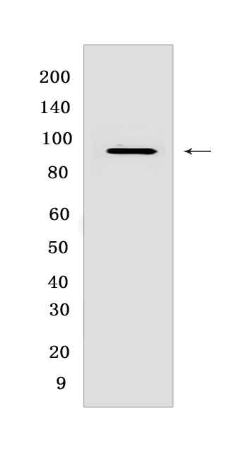
Western blot(SDS PAGE) analysis of extracts from Neuro-2a cells.Using PIP5K1CRabbit mAb [V8U8] at dilution of 1:1000 incubated at 4℃ over night.
Product information
Protein names :PIP5K1C,KIAA0589,PI51C_HUMAN,Phosphatidylinositol 4-phosphate 5-kinase type-1 gamma P-5-kinase 1 gamma)
UniProtID :O60331
MASS(da) :73,260
MW(kDa) :86 kDa
Form :Liquid
Purification :Protein A purification
Host :Rabbit
Isotype :IgG
sensitivity :Endogenous
Reactivity :Human,Mouse,Rat
- ApplicationDilution
- 免疫印迹(WB)1:1000-2000
- The optimal dilutions should be determined by the end user
Specificity :Antibody is produced by immunizing animals with a synthetic peptide at the sequence of human PIP5K1C
Storage :Antibody store in 10 mM PBS, 0.5mg/ml BSA, 50% glycerol. Shipped at 4°C. Store at-20°C or -80°C. Products are valid for one natural year of receipt.Avoid repeated freeze / thaw cycles.
WB Positive detected :Neuro-2a cells
Function : Catalyzes the phosphorylation of phosphatidylinositol 4-phosphate (PtdIns(4)P/PI4P) to form phosphatidylinositol 4,5-bisphosphate (PtdIns(4,5)P2/PIP2), a lipid second messenger that regulates several cellular processes such as signal transduction, vesicle trafficking, actin cytoskeleton dynamics, cell adhesion, and cell motility (PubMed:12422219, PubMed:22942276). PtdIns(4,5)P2 can directly act as a second messenger or can be utilized as a precursor to generate other second messengers: inositol 1,4,5-trisphosphate (IP3), diacylglycerol (DAG) or phosphatidylinositol-3,4,5-trisphosphate (PtdIns(3,4,5)P3/PIP3) (Probable). PIP5K1A-mediated phosphorylation of PtdIns(4)P is the predominant pathway for PtdIns(4,5)P2 synthesis (By similarity). Together with PIP5K1A, is required for phagocytosis, both enzymes regulating different types of actin remodeling at sequential steps (By similarity). Promotes particle attachment by generating the pool of PtdIns(4,5)P2 that induces controlled actin depolymerization to facilitate Fc-gamma-R clustering. Mediates RAC1-dependent reorganization of actin filaments. Required for synaptic vesicle transport (By similarity). Controls the plasma membrane pool of PtdIns(4,5)P2 implicated in synaptic vesicle endocytosis and exocytosis (PubMed:12847086). Plays a role in endocytosis mediated by clathrin and AP-2 (adaptor protein complex 2) (PubMed:12847086). Required for clathrin-coated pits assembly at the synapse (PubMed:17261850). Participates in cell junction assembly (PubMed:17261850). Modulates adherens junctions formation by facilitating CDH1/cadherin trafficking (PubMed:17261850). Required for focal adhesion dynamics. Modulates the targeting of talins (TLN1 and TLN2) to the plasma membrane and their efficient assembly into focal adhesions (PubMed:12422219). Regulates the interaction between talins (TLN1 and TLN2) and beta-integrins (PubMed:12422219). Required for uropodium formation and retraction of the cell rear during directed migration (By similarity). Has a role in growth factor-stimulated directional cell migration and adhesion (By similarity). Required for talin assembly into nascent adhesions forming at the leading edge toward the direction of the growth factor (PubMed:17635937). Negative regulator of T-cell activation and adhesion (By similarity). Negatively regulates integrin alpha-L/beta-2 (LFA-1) polarization and adhesion induced by T-cell receptor (By similarity). Together with PIP5K1A has a role during embryogenesis and together with PIP5K1B may have a role immediately after birth (By similarity)..
Tissue specificity :[Isoform 1]: Isoform 1 is strongly expressed in brain and also detected in heart and lung..,[Isoform 2]: Isoform 2 is strongly expressed in pancreas and liver and in lesser quantities in brain, heart, lung and kidney..,[Isoform 3]: Isoform 3 is detected in large amounts in heart and large intestine, is also present in lung, pancreas and thyroid, and to a lesser extent in brain, stomach and kidney..
Subcellular locationi :Cell membrane,Peripheral membrane protein,Cytoplasmic side. Endomembrane system. Cytoplasm. Cell junction, focal adhesion. Cell junction, adherens junction. Cell projection, ruffle membrane. Cell projection, phagocytic cup. Cell projection, uropodium.
IMPORTANT: For western blots, incubate membrane with diluted primary antibody in 1% w/v BSA, 1X TBST at 4°C overnight.
-
About
-
Product
-
Law
-
Literature
-

WeChat scan code follow us