
Homer1 Rabbit mAb [P268]Cat NO.: A49286
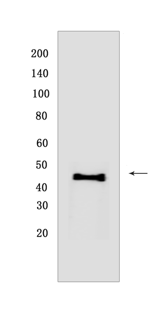
Western blot(SDS PAGE) analysis of extracts from HepG2 cells .Using Homer1Rabbit mAb [P268] at dilution of 1:1000 incubated at 4℃ over night.
Product information
Protein names :HOMER1,SYN47,HOME1_HUMAN,Homer protein homolog 1
UniProtID :Q86YM7
MASS(da) :40,277
MW(kDa) :46-48kDa
Form :Liquid
Purification :Protein A purification
Host :Rabbit
Isotype :IgG
sensitivity :Endogenous
Reactivity :Human,Mouse,Rat
- ApplicationDilution
- 免疫印迹(WB)1:1000-2000
- 免疫组化(IHC)1:100
- The optimal dilutions should be determined by the end user
Specificity :Antibody is produced by immunizing animals with a synthetic peptide at the sequence of human Homer1
Storage :Antibody store in 10 mM PBS, 0.5mg/ml BSA, 50% glycerol. Shipped at 4°C. Store at-20°C or -80°C. Products are valid for one natural year of receipt.Avoid repeated freeze / thaw cycles.
WB Positive detected :HepG2 cells
Function : Postsynaptic density scaffolding protein. Binds and cross-links cytoplasmic regions of GRM1, GRM5, ITPR1, DNM3, RYR1, RYR2, SHANK1 and SHANK3. By physically linking GRM1 and GRM5 with ER-associated ITPR1 receptors, it aids the coupling of surface receptors to intracellular calcium release. May also couple GRM1 to PI3 kinase through its interaction with AGAP2. Isoform 1 regulates the trafficking and surface expression of GRM5. Isoform 3 acts as a natural dominant negative, in dynamic competition with constitutively expressed isoform 1 to regulate synaptic metabotropic glutamate function. Isoform 3, may be involved in the structural changes that occur at synapses during long-lasting neuronal plasticity and development. Forms a high-order complex with SHANK1, which in turn is necessary for the structural and functional integrity of dendritic spines (By similarity). Negatively regulates T cell activation by inhibiting the calcineurin-NFAT pathway. Acts by competing with calcineurin/PPP3CA for NFAT protein binding, hence preventing NFAT activation by PPP3CA (PubMed:18218901)..
Subcellular locationi :Cytoplasm. Cell junction, synapse, postsynaptic density. Cell junction, synapse. Cell projection, dendritic spine.
IMPORTANT: For western blots, incubate membrane with diluted primary antibody in 1% w/v BSA, 1X TBST at 4°C overnight.
-
About
-
Product
-
Law
-
Literature
-

WeChat scan code follow us