
NADPH oxidase 4 Rabbit mAb [8K9S]Cat NO.: A16626
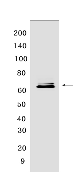
Western blot(SDS PAGE) analysis of extracts from human fetal kidney.Using NADPH oxidase 4Rabbit mAb [8K9S] at dilution of 1:1000 incubated at 4℃ over night.
Product information
Protein names :NOX4,RENOX,NOX4_HUMAN,NADPH oxidase 4 H-oxidase)
UniProtID :Q9NPH5
MASS(da) :66,932
MW(kDa) :63 kDa
Form :Liquid
Purification :Protein A purification
Host :Rabbit
Isotype :IgG
sensitivity :Endogenous
Reactivity :Human
- ApplicationDilution
- 免疫印迹(WB)1:1000-2000
- 免疫组化(IHC)1:100
- 免疫荧光(ICC/IF) 1:100
- The optimal dilutions should be determined by the end user
Specificity :Antibody is produced by immunizing animals with a synthetic peptide at the sequence of human NADPH oxidase 4
Storage :Antibody store in 10 mM PBS, 0.5mg/ml BSA, 50% glycerol. Shipped at 4°C. Store at-20°C or -80°C. Products are valid for one natural year of receipt.Avoid repeated freeze / thaw cycles.
WB Positive detected :human fetal kidney
Function : Constitutive NADPH oxidase which generates superoxide intracellularly upon formation of a complex with CYBA/p22phox. Regulates signaling cascades probably through phosphatases inhibition. May function as an oxygen sensor regulating the KCNK3/TASK-1 potassium channel and HIF1A activity. May regulate insulin signaling cascade. May play a role in apoptosis, bone resorption and lipolysaccharide-mediated activation of NFKB. May produce superoxide in the nucleus and play a role in regulating gene expression upon cell stimulation. Isoform 3 is not functional. Isoform 5 and isoform 6 display reduced activity., [Isoform 4]: Involved in redox signaling in vascular cells. Constitutively and NADPH-dependently generates reactive oxygen species (ROS). Modulates the nuclear activation of ERK1/2 and the ELK1 transcription factor, and is capable of inducing nuclear DNA damage. Displays an increased activity relative to isoform 1.
Tissue specificity :Expressed by distal tubular cells in kidney cortex and in endothelial cells (at protein level). Widely expressed. Strongly expressed in kidney and to a lower extent in heart, adipocytes, hepatoma, endothelial cells, skeletal muscle, brain, several brain tumor cell lines and airway epithelial cells..
Subcellular locationi :Endoplasmic reticulum membrane,Multi-pass membrane protein. Cell membrane,Multi-pass membrane protein. Cell junction, focal adhesion.
IMPORTANT: For western blots, incubate membrane with diluted primary antibody in 1% w/v BSA, 1X TBST at 4°C overnight.
-
About
-
Product
-
Law
-
Literature
-

WeChat scan code follow us