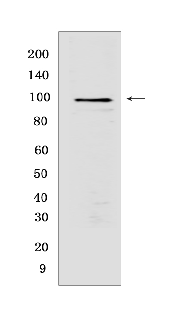
TPX2 Rabbit mAb [69U7]Cat NO.: A63882
Western blot(SDS PAGE) analysis of extracts from HEK-293 cells.Using TPX2Rabbit mAb [69U7] at dilution of 1:1000 incubated at 4℃ over night.
Product information
Protein names :TPX2,C20orf1,C20orf2,DIL2,HCA519,TPX2_HUMAN,Targeting protein for Xklp2
UniProtID :Q9ULW0
MASS(da) :85,653
MW(kDa) :100 kDa
Form :Liquid
Purification :Protein A purification
Host :Rabbit
Isotype :IgG
sensitivity :Endogenous
Reactivity :Human,Mouse,Rat
- ApplicationDilution
- 免疫印迹(WB)1:1000-2000
- 免疫组化(IHC)1:100
- 免疫荧光(ICC/IF) 1:100
- The optimal dilutions should be determined by the end user
Specificity :Antibody is produced by immunizing animals with a synthetic peptide at the sequence of human TPX2
Storage :Antibody store in 10 mM PBS, 0.5mg/ml BSA, 50% glycerol. Shipped at 4°C. Store at-20°C or -80°C. Products are valid for one natural year of receipt.Avoid repeated freeze / thaw cycles.
WB Positive detected :HEK-293 cells
Function : Spindle assembly factor required for normal assembly of mitotic spindles. Required for normal assembly of microtubules during apoptosis. Required for chromatin and/or kinetochore dependent microtubule nucleation. Mediates AURKA localization to spindle microtubules (PubMed:18663142, PubMed:19208764). Activates AURKA by promoting its autophosphorylation at 'Thr-288' and protects this residue against dephosphorylation (PubMed:18663142, PubMed:19208764). TPX2 is inactivated upon binding to importin-alpha (PubMed:26165940). At the onset of mitosis, GOLGA2 interacts with importin-alpha, liberating TPX2 from importin-alpha, allowing TPX2 to activates AURKA kinase and stimulates local microtubule nucleation (PubMed:26165940)..
Tissue specificity :Expressed in lung carcinoma cell lines but not in normal lung tissues.
Subcellular locationi :Nucleus. Cytoplasm, cytoskeleton, spindle. Cytoplasm, cytoskeleton, spindle pole.
IMPORTANT: For western blots, incubate membrane with diluted primary antibody in 1% w/v BSA, 1X TBST at 4°C overnight.


