MAGEC2 Rabbit mAb [PN38]Cat NO.: A20102
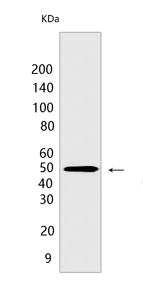
Western blot(SDS PAGE) analysis of extracts from A-375 cells .Using MAGEC2Rabbit mAb [PN38] at dilution of 1:1000 incubated at 4℃ over night.
Product information
Protein names :MAGEC2,HCA587,MAGEE1,MAGC2_HUMAN,Melanoma-associated antigen C2
UniProtID :Q9UBF1
MASS(da) :41,163
MW(kDa) :51 kDa
Form :Liquid
Purification :Protein A purification
Host :Rabbit
Isotype :IgG
sensitivity :Endogenous
Reactivity :Human,Mouse
- ApplicationDilution
- 免疫印迹(WB)1:1000-2000
- 免疫组化(IHC)1:100
- 免疫荧光(ICC/IF) 1:100
- The optimal dilutions should be determined by the end user
Specificity :Antibody is produced by immunizing animals with a synthetic peptide at the sequence of human MAGEC2
Storage :Antibody store in 10 mM PBS, 0.5mg/ml BSA, 50% glycerol. Shipped at 4°C. Store at-20°C or -80°C. Products are valid for one natural year of receipt.Avoid repeated freeze / thaw cycles.
WB Positive detected :A-375 cells
Function : Proposed to enhance ubiquitin ligase activity of RING-type zinc finger-containing E3 ubiquitin-protein ligases. In vitro enhances ubiquitin ligase activity of TRIM28 and stimulates p53/TP53 ubiquitination in presence of Ubl-conjugating enzyme UBE2H leading to p53/TP53 degradation. Proposed to act through recruitment and/or stabilization of the Ubl-conjugating enzymes (E2) at the E3:substrate complex..
Tissue specificity :Not expressed in normal tissues, except in germ cells in the seminiferous tubules and in Purkinje cells of the cerebellum. Expressed in various tumors, including melanoma, lymphoma, as well as pancreatic cancer, mammary gland cancer, non-small cell lung cancer and liver cancer. In hepatocellular carcinoma, there is an inverse correlation between tumor differentiation and protein expression, i.e. the lower the differentiation, the higher percentage of expression..
Subcellular locationi :Cytoplasm. Nucleus.
IMPORTANT: For western blots, incubate membrane with diluted primary antibody in 1% w/v BSA, 1X TBST at 4°C overnight.


