
Semaphorin 7a Rabbit mAb [1368]Cat NO.: A41857
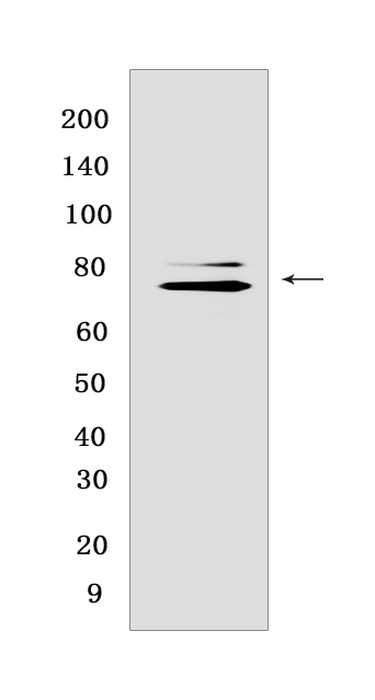
Western blot(SDS PAGE) analysis of extracts from human testis lysate.Using Semaphorin 7aRabbit mAb [1368] at dilution of 1:1000 incubated at 4℃ over night.
Product information
Protein names :SEMA7A,CD108,SEMAL,SEM7A_HUMAN,Semaphorin-7A
UniProtID :O75326
MASS(da) :74,824
MW(kDa) :75 kDa
Form :Liquid
Purification :Protein A purification
Host :Rabbit
Isotype :IgG
sensitivity :Endogenous
Reactivity :Human
- ApplicationDilution
- 免疫印迹(WB)1:1000-2000
- 免疫组化(IHC)1:100
- The optimal dilutions should be determined by the end user
Specificity :Antibody is produced by immunizing animals with a synthetic peptide at the sequence of human Semaphorin 7a
Storage :Antibody store in 10 mM PBS, 0.5mg/ml BSA, 50% glycerol. Shipped at 4°C. Store at-20°C or -80°C. Products are valid for one natural year of receipt.Avoid repeated freeze / thaw cycles.
WB Positive detected :human testis lysate
Function : Plays an important role in integrin-mediated signaling and functions both in regulating cell migration and immune responses. Promotes formation of focal adhesion complexes, activation of the protein kinase PTK2/FAK1 and subsequent phosphorylation of MAPK1 and MAPK3. Promotes production of pro-inflammatory cytokines by monocytes and macrophages. Plays an important role in modulating inflammation and T-cell-mediated immune responses. Promotes axon growth in the embryonic olfactory bulb. Promotes attachment, spreading and dendrite outgrowth in melanocytes..
Tissue specificity :Detected in skin keratinocytes and on endothelial cells from skin blood vessels (at protein level). Expressed in fibroblasts, keratinocytes, melanocytes, placenta, testis, ovary, spleen, brain, spinal chord, lung, heart, adrenal gland, lymph nodes, thymus, intestine and kidney..
Subcellular locationi :Cell membrane,Lipid-anchor, GPI-anchor,Extracellular side.
IMPORTANT: For western blots, incubate membrane with diluted primary antibody in 1% w/v BSA, 1X TBST at 4°C overnight.
-
About
-
Product
-
Law
-
Literature
-

WeChat scan code follow us