
EphA7 Rabbit mAb [3ZG2]Cat NO.: A74989
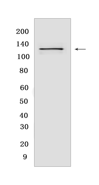
Western blot(SDS PAGE) analysis of extracts from LNCaP cells.Using EphA7 Rabbit mAb [3ZG2] at dilution of 1:1000 incubated at 4℃ over night.
Product information
Protein names :EPHA7,EHK3,HEK11,EPHA7_HUMAN,Ephrin type-A receptor 7
UniProtID :Q15375
MASS(da) :112,097
MW(kDa) :130 kDa
Form :Liquid
Purification :Protein A purification
Host :Rabbit
Isotype :IgG
sensitivity :Endogenous
Reactivity :Human
- ApplicationDilution
- 免疫印迹(WB)1:1000-2000
- The optimal dilutions should be determined by the end user
Specificity :Antibody is produced by immunizing animals with a synthetic peptide at the sequence of Human EphA7
Storage :Antibody store in 10 mM PBS, 0.5mg/ml BSA, 50% glycerol. Shipped at 4°C. Store at-20°C or -80°C. Products are valid for one natural year of receipt.Avoid repeated freeze / thaw cycles.
WB Positive detected :LNCaP cells
Function : Receptor tyrosine kinase which binds promiscuously GPI-anchored ephrin-A family ligands residing on adjacent cells, leading to contact-dependent bidirectional signaling into neighboring cells. The signaling pathway downstream of the receptor is referred to as forward signaling while the signaling pathway downstream of the ephrin ligand is referred to as reverse signaling. Among GPI-anchored ephrin-A ligands, EFNA5 is a cognate/functional ligand for EPHA7 and their interaction regulates brain development modulating cell-cell adhesion and repulsion. Has a repellent activity on axons and is for instance involved in the guidance of corticothalamic axons and in the proper topographic mapping of retinal axons to the colliculus. May also regulate brain development through a caspase(CASP3)-dependent proapoptotic activity. Forward signaling may result in activation of components of the ERK signaling pathway including MAP2K1, MAP2K2, MAPK1 AND MAPK3 which are phosphorylated upon activation of EPHA7..
Tissue specificity :Widely expressed.
Subcellular locationi :Cell membrane,Single-pass type I membrane protein.
IMPORTANT: For western blots, incubate membrane with diluted primary antibody in 1% w/v BSA, 1X TBST at 4°C overnight.
-
About
-
Product
-
Law
-
Literature
-

WeChat scan code follow us