
Eps8 Rabbit mAb [X30Q]Cat NO.: A69305
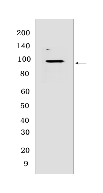
Western blot(SDS PAGE) analysis of extracts from LOVO cells.Using Eps8 Rabbit mAb [X30Q] at dilution of 1:1000 incubated at 4℃ over night.
Product information
Protein names :EPS8,EPS8_HUMAN,Epidermal growth factor receptor kinase substrate 8
UniProtID :Q12929
MASS(da) :91,882
MW(kDa) :95 kDa
Form :Liquid
Purification :Protein A purification
Host :Rabbit
Isotype :IgG
sensitivity :Endogenous
Reactivity :Human,Mouse,Rat
- ApplicationDilution
- 免疫印迹(WB)1:1000-2000
- The optimal dilutions should be determined by the end user
Specificity :Antibody is produced by immunizing animals with a synthetic peptide at the sequence of Human Eps8
Storage :Antibody store in 10 mM PBS, 0.5mg/ml BSA, 50% glycerol. Shipped at 4°C. Store at-20°C or -80°C. Products are valid for one natural year of receipt.Avoid repeated freeze / thaw cycles.
WB Positive detected :LOVO cells
Function : Signaling adapter that controls various cellular protrusions by regulating actin cytoskeleton dynamics and architecture. Depending on its association with other signal transducers, can regulate different processes. Together with SOS1 and ABI1, forms a trimeric complex that participates in transduction of signals from Ras to Rac by activating the Rac-specific guanine nucleotide exchange factor (GEF) activity. Acts as a direct regulator of actin dynamics by binding actin filaments and has both barbed-end actin filament capping and actin bundling activities depending on the context. Displays barbed-end actin capping activity when associated with ABI1, thereby regulating actin-based motility process: capping activity is auto-inhibited and inhibition is relieved upon ABI1 interaction. Also shows actin bundling activity when associated with BAIAP2, enhancing BAIAP2-dependent membrane extensions and promoting filopodial protrusions. Involved in the regulation of processes such as axonal filopodia growth, stereocilia length, dendritic cell migration and cancer cell migration and invasion. Acts as a regulator of axonal filopodia formation in neurons: in the absence of neurotrophic factors, negatively regulates axonal filopodia formation via actin-capping activity. In contrast, it is phosphorylated in the presence of BDNF leading to inhibition of its actin-capping activity and stimulation of filopodia formation. Component of a complex with WHRN and MYO15A that localizes at stereocilia tips and is required for elongation of the stereocilia actin core. Indirectly involved in cell cycle progression,its degradation following ubiquitination being required during G2 phase to promote cell shape changes..
Tissue specificity :Expressed in all tissues analyzed, including heart, brain, placenta, lung, liver, skeletal muscle, kidney and pancreas. Expressed in all epithelial and fibroblastic lines examined and in some, but not all, hematopoietic cells.
Subcellular locationi :Cytoplasm, cell cortex. Cell projection, ruffle membrane. Cell projection, growth cone. Cell projection, stereocilium. Cell junction, synapse, synaptosome.
IMPORTANT: For western blots, incubate membrane with diluted primary antibody in 1% w/v BSA, 1X TBST at 4°C overnight.
-
About
-
Product
-
Law
-
Literature
-

WeChat scan code follow us