
FAP Rabbit mAb [D151]Cat NO.: A48792
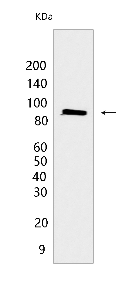
Western blot(SDS PAGE) analysis of extracts from M14 cells.Using FAP Rabbit mAb [D151] at dilution of 1:1000 incubated at 4℃ over night.
Product information
Protein names :FAP,SEPR_HUMAN,Prolyl endopeptidase FAP [Cleaved into: Antiplasmin-cleaving enzyme FAP, soluble form ]
UniProtID :Q12884
MASS(da) :87,713
MW(kDa) :90 kDa
Form :Liquid
Purification :Protein A purification
Host :Rabbit
Isotype :IgG
sensitivity :Endogenous
Reactivity :Human
- ApplicationDilution
- 免疫印迹(WB)1:1000-2000
- The optimal dilutions should be determined by the end user
Specificity :Antibody is produced by immunizing animals with a synthetic peptide at the sequence of Human FAP
Storage :Antibody store in 10 mM PBS, 0.5mg/ml BSA, 50% glycerol. Shipped at 4°C. Store at-20°C or -80°C. Products are valid for one natural year of receipt.Avoid repeated freeze / thaw cycles.
WB Positive detected :M14 cells
Function : Cell surface glycoprotein serine protease that participates in extracellular matrix degradation and involved in many cellular processes including tissue remodeling, fibrosis, wound healing, inflammation and tumor growth. Both plasma membrane and soluble forms exhibit post-proline cleaving endopeptidase activity, with a marked preference for Ala/Ser-Gly-Pro-Ser/Asn/Ala consensus sequences, on substrate such as alpha-2-antiplasmin SERPINF2 and SPRY2 (PubMed:14751930, PubMed:16223769, PubMed:16480718, PubMed:16410248, PubMed:17381073, PubMed:18095711, PubMed:21288888, PubMed:24371721). Degrade also gelatin, heat-denatured type I collagen, but not native collagen type I and IV, vitronectin, tenascin, laminin, fibronectin, fibrin or casein (PubMed:9065413, PubMed:2172980, PubMed:7923219, PubMed:10347120, PubMed:10455171, PubMed:12376466, PubMed:16223769, PubMed:16651416, PubMed:18095711). Also has dipeptidyl peptidase activity, exhibiting the ability to hydrolyze the prolyl bond two residues from the N-terminus of synthetic dipeptide substrates provided that the penultimate residue is proline, with a preference for Ala-Pro, Ile-Pro, Gly-Pro, Arg-Pro and Pro-Pro (PubMed:10347120, PubMed:10593948, PubMed:16175601, PubMed:16223769, PubMed:16651416, PubMed:16410248, PubMed:17381073, PubMed:21314817, PubMed:24371721, PubMed:24717288). Natural neuropeptide hormones for dipeptidyl peptidase are the neuropeptide Y (NPY), peptide YY (PYY), substance P (TAC1) and brain natriuretic peptide 32 (NPPB) (PubMed:21314817). The plasma membrane form, in association with either DPP4, PLAUR or integrins, is involved in the pericellular proteolysis of the extracellular matrix (ECM), and hence promotes cell adhesion, migration and invasion through the ECM. Plays a role in tissue remodeling during development and wound healing. Participates in the cell invasiveness towards the ECM in malignant melanoma cancers. Enhances tumor growth progression by increasing angiogenesis, collagen fiber degradation and apoptosis and by reducing antitumor response of the immune system. Promotes glioma cell invasion through the brain parenchyma by degrading the proteoglycan brevican. Acts as a tumor suppressor in melanocytic cells through regulation of cell proliferation and survival in a serine protease activity-independent manner..
Tissue specificity :Expressed in adipose tissue. Expressed in the dermal fibroblasts in the fetal skin. Expressed in the granulation tissue of healing wounds and on reactive stromal fibroblast in epithelial cancers. Expressed in activated fibroblast-like synoviocytes from inflamed synovial tissues. Expressed in activated hepatic stellate cells (HSC) and myofibroblasts from cirrhotic liver, but not detected in normal liver. Expressed in glioma cells (at protein level). Expressed in glioblastomas and glioma cells. Isoform 1 and isoform 2 are expressed in melanoma, carcinoma and fibroblast cell lines..
Subcellular locationi :[Prolyl endopeptidase FAP]: Cell surface. Cell membrane,Single-pass type II membrane protein. Cell projection, lamellipodium membrane,Single-pass type II membrane protein. Cell projection, invadopodium membrane,Single-pass type II membrane protein. Cell projection, ruffle membrane,Single-pass type II membrane protein. Membrane,Single-pass type II membrane protein.
IMPORTANT: For western blots, incubate membrane with diluted primary antibody in 1% w/v BSA, 1X TBST at 4°C overnight.
-
About
-
Product
-
Law
-
Literature
-

WeChat scan code follow us