CD55 Rabbit mAb[3VWR]Cat NO.: A22566
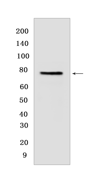
Western blot(SDS PAGE) analysis of extracts from HepG2 cells.Using CD55 Rabbit mAb IgG [3VWR] at dilution of 1:1000 incubated at 4℃ over night.
Product information
Protein names :CD55,CR,DAF,DAF_HUMAN,Complement decay-accelerating factor
UniProtID :P08174
MASS(da) :41,400
MW(kDa) :78KDa
Form :Liquid
Purification :Protein A purification
Host :Rabbit
Isotype :IgG
sensitivity :Endogenous
Reactivity :Human
- ApplicationDilution
- 免疫印迹(WB)1:1000-2000,
- 免疫组化(IHC)1:100
- The optimal dilutions should be determined by the end user
Specificity :Antibody is produced by immunizing animals with a synthetic peptide of human CD55.
Storage :Antibody store in 10 mM PBS, 0.5mg/ml BSA, 50% glycerol. Shipped at 4°C. Store at-20°C or -80°C. Products are valid for one natural year of receipt.Avoid repeated freeze / thaw cycles.
WB Positive detected :HepG2 cells
Function : This protein recognizes C4b and C3b fragments that condense with cell-surface hydroxyl or amino groups when nascent C4b and C3b are locally generated during C4 and c3 activation. Interaction of daf with cell-associated C4b and C3b polypeptides interferes with their ability to catalyze the conversion of C2 and factor B to enzymatically active C2a and Bb and thereby prevents the formation of C4b2a and C3bBb, the amplification convertases of the complement cascade (PubMed:7525274). Inhibits complement activation by destabilizing and preventing the formation of C3 and C5 convertases, which prevents complement damage (PubMed:28657829).., (Microbial infection) Acts as a receptor for Coxsackievirus A21, coxsackieviruses B1, B3 and B5.., (Microbial infection) Acts as a receptor for Human enterovirus 70 and D68 (Probable).., (Microbial infection) Acts as a receptor for Human echoviruses 6, 7, 11, 12, 20 and 21..
Tissue specificity :Expressed on the plasma membranes of all cell types that are in intimate contact with plasma complement proteins. It is also found on the surfaces of epithelial cells lining extracellular compartments, and variants of the molecule are present in body fluids and in extracellular matrix.
Subcellular locationi :[Isoform 1]: Cell membrane,Single-pass type I membrane protein.,[Isoform 2]: Cell membrane,Lipid-anchor, GPI-anchor.,[Isoform 3]: Secreted.,[Isoform 4]: Secreted.,[Isoform 5]: Secreted.,[Isoform 6]: Cell membrane,Lipid-anchor, GPI-anchor.,[Isoform 7]: Cell membrane,Lipid-anchor, GPI-anchor.
IMPORTANT: For western blots, incubate membrane with diluted primary antibody in 1% w/v BSA, 1X TBST at 4°C overnight.


