
Ephrin B2 Rabbit mAb[T3T2]Cat NO.: A87714
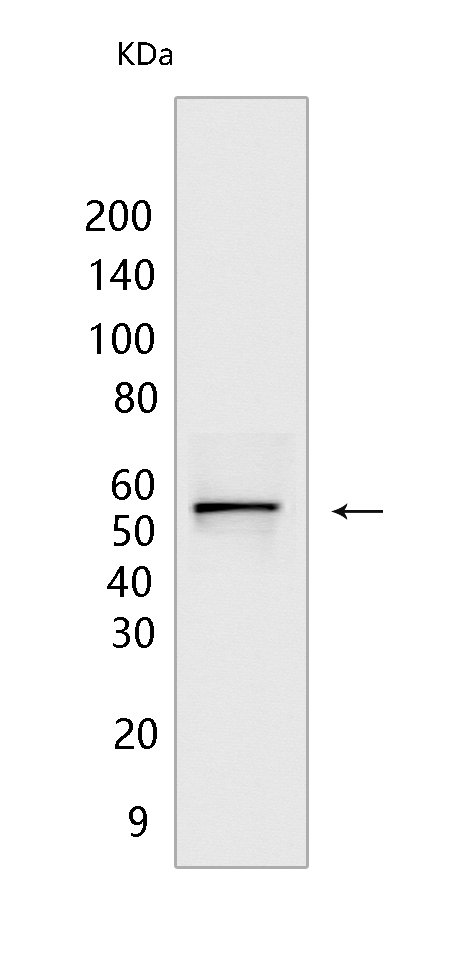
Western blot(SDS PAGE) analysis of extracts from 293T cells.Using Ephrin B2 Rabbit mAb IgG [T3T2] at dilution of 1:1000 incubated at 4℃ over night.
Product information
Protein names :EFNB2,EPLG5,HTKL,LERK5,EFNB2_HUMAN,Ephrin-B2
UniProtID :P52799
MASS(da) :36,923
MW(kDa) :55KDa
Form :Liquid
Purification :Protein A purification
Host :Rabbit
Isotype :IgG
sensitivity :Endogenous
Reactivity :Human,Mouse,Rat
- ApplicationDilution
- 免疫印迹(WB)1:1000-2000,
- 免疫组化(IHC)1:100
- The optimal dilutions should be determined by the end user
Specificity :Antibody is produced by immunizing animals with a synthetic peptide of human Ephrin B2.
Storage :Antibody store in 10 mM PBS, 0.5mg/ml BSA, 50% glycerol. Shipped at 4°C. Store at-20°C or -80°C. Products are valid for one natural year of receipt.Avoid repeated freeze / thaw cycles.
WB Positive detected :293T cells
Function : Cell surface transmembrane ligand for Eph receptors, a family of receptor tyrosine kinases which are crucial for migration, repulsion and adhesion during neuronal, vascular and epithelial development. Binds promiscuously Eph receptors residing on adjacent cells, leading to contact-dependent bidirectional signaling into neighboring cells. The signaling pathway downstream of the receptor is referred to as forward signaling while the signaling pathway downstream of the ephrin ligand is referred to as reverse signaling. Binds to receptor tyrosine kinase including EPHA4, EPHA3 and EPHB4. Together with EPHB4 plays a central role in heart morphogenesis and angiogenesis through regulation of cell adhesion and cell migration. EPHB4-mediated forward signaling controls cellular repulsion and segregation from EFNB2-expressing cells. May play a role in constraining the orientation of longitudinally projecting axons.., (Microbial infection) Acts as a receptor for Hendra virus and Nipah virus..
Tissue specificity :Lung and kidney.
Subcellular locationi :Cell membrane,Single-pass type I membrane protein. Cell junction, adherens junction.
IMPORTANT: For western blots, incubate membrane with diluted primary antibody in 1% w/v BSA, 1X TBST at 4°C overnight.
-
About
-
Product
-
Law
-
Literature
-

WeChat scan code follow us