FGF1 Rabbit mAb[Q4SE]Cat NO.: A95952
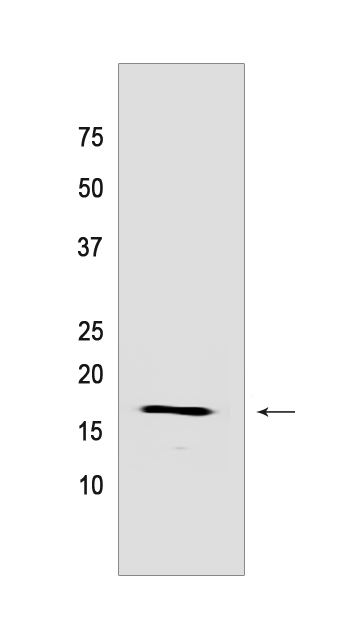
Western blot(SDS PAGE) analysis of extracts from Mouse kidney tissue lysate.Using FGF1 Rabbit mAb IgG [Q4SE] at dilution of 1:1000 incubated at 4℃ over night.
Product information
Protein names :FGF1,FGFA,FGF1_HUMAN,Fibroblast growth factor 1
UniProtID :P05230
MASS(da) :17,460
MW(kDa) :17KDa
Form :Liquid
Purification :Protein A purification
Host :Rabbit
Isotype :IgG
sensitivity :Endogenous
Reactivity :Human,Mouse,Rat
- ApplicationDilution
- 免疫印迹(WB)1:1000-2000,
- 免疫荧光(ICC/IF) 1:100
- The optimal dilutions should be determined by the end user
Specificity :Antibody is produced by immunizing animals with a synthetic peptide of human FGF1.
Storage :Antibody store in 10 mM PBS, 0.5mg/ml BSA, 50% glycerol. Shipped at 4°C. Store at-20°C or -80°C. Products are valid for one natural year of receipt.Avoid repeated freeze / thaw cycles.
WB Positive detected :Mouse kidney tissue lysate
Function : Plays an important role in the regulation of cell survival, cell division, angiogenesis, cell differentiation and cell migration. Functions as potent mitogen in vitro. Acts as a ligand for FGFR1 and integrins. Binds to FGFR1 in the presence of heparin leading to FGFR1 dimerization and activation via sequential autophosphorylation on tyrosine residues which act as docking sites for interacting proteins, leading to the activation of several signaling cascades. Binds to integrin ITGAV:ITGB3. Its binding to integrin, subsequent ternary complex formation with integrin and FGFR1, and the recruitment of PTPN11 to the complex are essential for FGF1 signaling. Induces the phosphorylation and activation of FGFR1, FRS2, MAPK3/ERK1, MAPK1/ERK2 and AKT1 (PubMed:18441324, PubMed:20422052). Can induce angiogenesis (PubMed:23469107)..
Tissue specificity :Predominantly expressed in kidney and brain. Detected at much lower levels in heart and skeletal muscle..
Subcellular locationi :Secreted. Cytoplasm. Cytoplasm, cell cortex. Cytoplasm, cytosol. Nucleus.
IMPORTANT: For western blots, incubate membrane with diluted primary antibody in 1% w/v BSA, 1X TBST at 4°C overnight.


